Muscular tissues
Category : 11th Class
Contractility and motility (movement) are fundamental properties of protoplasm. That is why, all cells possess potential motility. Contraction for motility in the cells results essentially from the interaction of two contractile proteins, actin and myosin. These tissues are obviously responsible for movements of organs and locomotion of the body in response to stimuli. These develop from embryonic mesoderm except for those of the iris and ciliary body of eyes, which are ectodermal in origin. About 40% to 50% of our body mass is of muscles. The muscle cells are always elongated, slender and spindle-shaped, fibre-like cells, These are, therefore called muscle fibres. These possess large numbers of myofibrils formed of actin and myosin. Muscle cells lose capacity to divide, multiply and regenerate to a great extent. Study of muscle is called myology. Types of muscle are following –
Striated or striped muscles
Most muscles of body are striated. These generally bring about voluntary movements under conscious control of brain and, hence, called voluntary muscles. Most of these are inserted at both ends upon bones in different parts of the body depend upon these muscles. Hence, these are also called skeletal muscles. Movements of limbs and the body solely depend upon these muscles. Hence these are also called somatic muscles. These are also called phasic type of muscles, because contraction in these is rapid, but brief and fatigue occurs quickly.
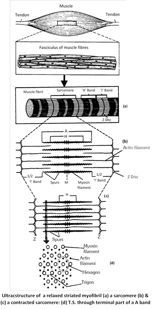
Fine structure of striated muscle fibres : Striated muscle fibres shows transverse striation in the form of regular alternate dark A (anisotropic) and light I (isotropic) bands. The ‘A’ band contains about \[120{AA}\] thick and \[1.8\,\,\mu \] long “myosin filaments”. The I band contains about \[60{AA}\] thick and \[1.0\,\,\mu \] long “actin filament” which are twice as many as myosin filaments. Each I band is divided into two equal halves by a thin, fibrous and transverse zig-zag partition, called ‘Z’ band (‘ Z’ disc) or Krause’s membrane. Each segment of a fibril between two adjacent ‘Z’ bands is called a sarcomere. It is \[2.3\,\,\mu \] long in uncontracted mammalian striated fibres. A slender transverse line, the ‘M’ or Hansen’s line is visible in middle of each ‘A’ band. The major, middle region of ‘A’ band is comparatively lighter, but its terminal parts appear darker. The middle lighter region is called ‘H’ zone. Due to the geomatric bonding pattern, the end of each myosin filament is, thus, encircled by the ends of six actin filaments (hexagon), while the end of each actin filaments is encircled by the ends of three myosin filaments (trigon).
Ultrastructure of myofilaments : At the molecular level, each myosin filament is composed of about 500 thread-like myosin molecules. Three different kinds of proteins participate in the composition of actin filaments. The major part of an actin filament is a coiled double helical strand whose each arm is a linear polymer of small and globular molecules (monomers) actin protein. Another coiled double helical, but thiner, strand runs along the whole length of actin strand. Each arm of this strand is a polymer of fibre-like molecules of tropomyosin protein. The third protein is troponin.
Working of striated muscles : H.E. Huxley and A.F. Huxley in 1954 proposed a theory to explain the process of muscular contraction. This theory is known as ‘sliding filament theory’. It was observed that when a fibril contracts, its ‘A’ bands remain intact, while the ‘I’ bands progressively shorten and eventually disappear when the fibril has shortened to about 65% of its resting length. At this stage. ‘H’ zones also disappear because the actin filaments of both sides in each sarcomere reach, and may even overlap each other at the “M” line, and the ‘Z’ lines now touch the ends of myosin filaments. It was further observed that if a fibre is mechanically streched, the zones of overlap between thick and thin filaments are shorter than in resting condition, resulting in wider ‘H’ zones. These observations led Huxley to propose that shortening of the fibrils in contraction is brought about by sliding movement of actin filaments over myosin filaments towards “M” line by means of rapidly forming and breaking cross bridges or rachets at the spurs of myosin filaments. Thus, the sarcomere were recognised as the ‘ultimate units of contraction’.
Smooth muscles
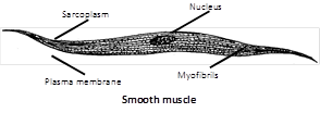
These are called smooth, plain nonstriated involuntary or unstriped muscles due to absence of striations. These occur in the walls of hollow internal organs (alimentary canal, gall bladder, bile ducts, respiratory tracts, uterus, urinogenital ducts, urinary bladder, blood vessels, etc.), in capsules of lymph glands, spleen etc., in iris and ciliary body of eyes, skin dermis, penis and other accessory genitalia, etc. There is no connection of these muscles with bones. Smooth muscles of skin dermis, called arrector pilli muscles, are associated with hair roots, and are responsible for flesh (erection of hairs). Those of penis form a muscular network which helps in its erection and limping.
Structure : Smooth muscle fibre is unbranched goose-spindle shaped, uninucleated and has no sarcolemma. Contraction is slow, involuntary under the control of ANS. Functionally smooth muscles are of two types –
(1) Single-unit smooth muscle : Single unit smooth muscle fibres are composed of muscle fibres closely joined together, contract as a single unit. e.g., urinary bladder, gastrointestinal tract, small arteries and small veins.
(2) Multi-unit smooth muscles : Are composed of more independent muscle fibres, contract as separate units e.g. – hair root muscle, muscles on the wall of large blood vessels, ciliary muscles, muscles of iris and bronchi.
Cardiac muscles
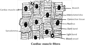
Heart wall (also the wall of large veins just where these enter into the heart) is made up of cardiac muscles and, hence, called myocardium. Structurally, these muscles resemble striated muscles but, functioning independently of the conscious control of brain, these are involuntary like the smooth muscles. Cardiac muscle cells of fibres are comparatively shorter and thicker, cylindrical, mostly uninucleate with a central nucleus, somewhat branched and covered by a sarcolemma.
Characteristics of a muscle
Antagonistic muscles : The striated muscles occur in antagonistic pairs; one pulls a bone in one direction, while the other pulls it back in reverse direction to its normal position. For example, the biceps muscle, extending from shoulder to radius, bends or flexes the arm at the elbow, whereas the triceps extending from ulna to the shoulder, straightens the arm. Thus, biceps is a flexor and triceps an extensor for bending the arm.
Single twitch : When a muscle receives a single excitation impulse, it respond by a sudden partial contraction (twitch) lasting for about 0.5 second in man. Each twitch is followed by a refractory period during which the muscle does not respond to next stimulus. The refractory period is, however, so short (0.002 second) that the muscle can respond to the second stimulus while still in contraction phase in response to the first stimulus.
Tetanus : Generally, whole muscles contract, not in a single twitch, but in sustained contractions evoked by a series of nerve impulses reaching them in rapid succession. Such a sustained contraction is called tetanus. Described above should not be confused with the disease of “tetanus” (lock jaw) caused by tetanus bacillus. This disease is characterised by abnormal muscular contractions. Nor it should be confused with “tetany” which is muscular spasm occurring due to deficiency of parathyroid hormone.
Muscle tone or “Tonus” : Even at rest the striated muscles normally remain in a state of mild sustained partial contraction to maintain the body posture. This is called muscle tone. It is a mild state of tetanus.
Paralysis : When supply of motor impulses to a muscle is completely cut off due to destruction, either of the control centres in brain, or of the concerned motor nerves, or due to blocking of myoneural junctions by the use of certain drugs, the muscle function is completely impaired. This is called paralysis of the muscle.
Muscle fatigue : A muscle that has contracted many times at short intervals, exhausts its store of ATP and glycogen and accumulates lactic acid. Hence its contractility gradually decreases and finally stops.
Oxygen debt : During active work or exercise, the rate of oxygen supply by the lungs falls short of the requirement of the muscles. Hence, lactic acid accumulates in the muscles and the breathing gradually becomes hard to enhance \[{{O}_{2}}\] intake by the lungs. This is called oxygen debt.
Involuntary action of skeletal muscles : Muscles are capable of utilizing, in their mechanical work, only about 20% to 40% of energy liberated from glucose. The unutilized energy is lost as “heat” dissipated into the environment. This heat helps in maintenance of body temperature. “Shivering with cold” in winter is caused by a quick involuntary reaction of striated muscles.
Rigor mortis : Rigidity that develops in the muscles after death is known as rigor mortis. It is due to permanent irreversible contraction, establishment of permanent link between actin and myosin and also fall in the concentration of ATP molecules.
Cori’s cycle : Lactic acid is transported by blood to liver and there it is converted to glycogen through Cori’s cycle.
Contraction period : Time taken in sliding of filament is called contraction time. (10 to 100 milli second).
Relaxation time : It is time taken in relaxation of fibre i.e. active transport of calcium from sarcoplasm to cisternae. (10 to 100 milli second)
Refractory period : It is time in a muscle or nerve fibre when they are non responding to second stimulus. Infact in this period there is temporary loss of excitability. Refractory period for skeletal and cardiac muscle is 5 and 300 milli second respectively.
Hypertrophy and Atrophy of muscles : Muscles which are put to excessive work become thick and strong. This is called their hypertrophy. Conversely, if certain muscles are not used for a long period, those become thin and weak. This is called their atrophy (disuse atrophy). Cardiac muscle have a poor regenrative power.


You need to login to perform this action.
You will be redirected in
3 sec

