Skeletal Tissue
Category : 11th Class
It provide support and surface for attachment of muscle. Skeletal connective tissue form the frame work of body. It provide rigidity to body. These protect the various organ and help in locomotion. It is of three types : Cartilage, Bones, Notochord.
Cartilage
Cartilage is a solid but semi-rigid and flexible connective tissue. Cartilage is a nonvascular connective tissue, consisting of cells embeded in a resilent matrix of chondrin. Chondrin is a protein of cartilage. Regeneration of cartilage can occur from its peri-chondrium. Cartilage is said to be metabolically nearly inactive. In kids the cartilage cells show 2 types of growth.
(1) Appositional or Perichondral or Secondary or Exogenous growth : It is due to deposition of matrix and division of chondrogenic cells of periphery. It leads to growth in thickness.
(2) Endogenous or Interstitial growth : It is due to deposition of matrix and division in inner cells of cartilage. It leads to growth in size.
Types of cartilage : It is of following types –
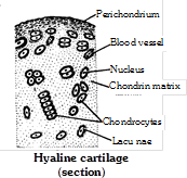
(1) Hyaline cartilage : It is most primitive and glass like cartilage. Its matrix is transparent homogenous and pearly white or bluish green in colour, contain chondrin. It is slightly elastic and also known as articular cartilage because it forms the articular surface of joints. Hyaline cartilage is found in trachea, larynx and bronchi, limb bones (called hyaline cap), sternum, in the hyoid apparatus nasal septum, ribs (sternal parts) larynx (cricoid, thyroid), nasal cartilage (nasal septum).
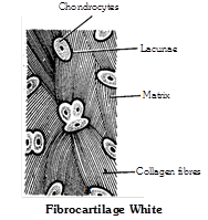
(2) Fibro cartilage (White fibrous cartilage) : In this cartilage, the small amount of matrix of cartilage is packed with large number of bundles of thick white (collagen) fibres. So it is toughest and less flexible. It is found in intervertebral discs and acts as shock absorber. It is also found in pubic symphysis and helps in parturition (child birth). The intervertebral discs remain contracted when the body is active, but relaxed when the body is at rest. That is why, our body becomes a bit taller during sleep and after death.
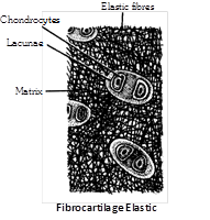
(3) Elastic cartilage (Yellow elastic cartilage) : In this cartilage, the matrix is packed with yellow or elastic fibres which run in all directions to form a network. Owing to the presence of yellow fibres, it is very flexible. It gives recoiling power to structures. It is found in mammalian pinna, pharyngotympanic tube, epiglottis, some laryngeal and bronchiolar cartilages.
(4) Calcified cartilage : It is modified hyaline cartilage, It is hard and non elastic due to deposition of calcium salt-hydroxy appetite in matrix. It is found in pubis of old frog, supra-scapula of frog, quadrate cartilage of frog, shark vertebrae, in man ends of long bone, head of humerus and femur. Calcification may also occur as a regular growth process of bone due to age. It reduces elasticity of the cartilage and makes it more rigid.
Bone
Bone is a highly calcified (mineralized), hard and rigid connective tissue. It is the major component of adult vertebrate endoskeleton. Besides its mechanical function of supporting the body architecture and internal organs as a frame work, of protecting delicate organ like brain, heart, etc. of forming to muscles to facilitate movement and locomotion, the bone is also a metabolically dynamic tissue which functions as a homeostatic reservoir of ions of calcium, magnesium, phosphorous, etc. About 97% of total calcium of body occurs in the endoskeleton.
Structure of bone
Periosteum : It is a membrane that forms an envelop around the bone. Periosteum is comprises of two distinct layers. Outer layer consist of thin white fibrous connective tissue. Inner layer consist of osteoblasts, osteoblasts are spider like bone cells, also known as bone forming cells, because they produces new bone materials.
Matrix : Matrix is composed of protein called ossein. The matrix forms thin plates called lamellae. Lamellae are of three types. Haversian lamellae (occur around Haversian canal) concentric or circumferential lamellae (inner to periosteum and outer to endosteum) and interstitial lamellae (between Haversian system). In the lamellae minute bone cells osteocytes are present.
Endosteum : It is present outer to the bone marrow cavity. Endosteum is a membrane which lines the marrow cavity. It is comprises of two distinct layers, one is of fibrous connective tissue and another is osteoblasts.

Bone marrow : Bone marrow is a specialized type of soft, diffuse connective tissue called “Myeloid tissue”. It takes part in production of blood cells hence known as haemopoietic tissue. It is composed of adipose tissue, areolar tissue and blood. It is of two types –
(1) Red bone marrow : Red in colour due to presence of lot of blood vessels. In foetal life and at birth present in entire skeleton. After 5th year red bone marrow replaced by yellow bone marrow, at 20-25 years red bone marrow present at ribs, sternum, clavicles, vertebrae, scapula, pelvis, epiphysis of humerus and femur. Produces RBCs, WBC, platelets, granular, leucocytes like basophils eosinophils and neutrophils.
(2) Yellow bone marrow : Yellow in colour and has much fatty tissue (adipose tissue), present in shaft of long bones. Produces blood cells in emergency i.e. at the time of excessive loss of blood, yellow bone marrow may be replaced by red bone marrow in anaemia.
Haversian system : A haversian canal, its lamellae and osteocytes form a haversian system. Haversian canals are found in bone matrix of long bone, like humerus of mammals. Haversian canals contain artery and veins, osteoblasts in areolar tissue, nerves and lymph. It is also called osteon.
Types of bone cells : Four types of cells are found in bone :
(1) Osteoprogenitor cell : Develops into osteoblast cell due to mitotic cell division.
(2) Osteoblast : Bone forming cells found in all bone surfaces. It is small cells synthesize and secrete osteoid, an important part of ground substance. Process of osteoblast is called canaliculi.
(3) Osteocyte : Mature, nondividing osteoblast surrounded by matrix, lying within lacunae.
(4) Osteoclast : Bone destroying cells take part in reabsorption of bones, contain large amount of acid phosphatase enzyme.
Types of bone
On the basis of their texture : The bones are divided into two categories spongy or cancellous or tubecular bones and compact or periosteal bones.
Differences between Spongy bone and Compact bone
|
Characters |
Spongy bone |
Compact bone |
|
Arrangement of lamellae |
There is no regular Haversian system so have spongy texture. |
Have regular Haversian system |
|
Occurrence |
In skull bones, ribs, centrum of vertebrae and epiphyses of long bones |
In the shaft (diaphysis) of long bones |
|
Marrow cavity |
Broad |
Narrow |
|
Type of bone marrow |
Red marrow in the spaces between lamellae |
Yellow marrow in marrow cavity |
|
Function |
Marrow forms RBCs and Granular WBCs, Platelets |
Marrow stores fats |
On the basis of origin of bone : Ossification or osteogenesis is the process of bone formation. A bones are classified into four categories – Cartilaginous, Dermal, Sesamoid and Visceral bones
Differences between Cartilaginous, Dermal, Sesamoid and Visceral bones
|
Cartilaginous (Endochondrial) bone |
Dermal (Intramembranous) bone |
Sesamoid bone |
Visceral bone |
|
These are formed by ossification directly on the cartilages and formation involves deposition of body matter by osteoblasts and resorption by osteoclast. |
These are formed by ossification in the dermis of the skin. |
These are formed by ossification at the joints of the bones or on the tendon and ligament. |
They are formed in the soft organs. |
|
These are elongated and hard bones Examples : Vertebrae, humerus, femur and fibula, girdles. |
These are membrane-like bones. Examples : skull bones, phalanges, clavicles. |
These are small sized disc like bones. Example : patella bone (knee cap). |
Examples : oscordis, ospenis, osclitoris. |
On the basis of treatment : These are of two types – Dried bone and Decalcified bone
Differences between Dried bone and Decalcified bone
|
Characters |
Dried bone |
Decalcified bone |
|
Type of treatment |
Subjected to high temperature. |
Subjected to dilute solution of HCl. |
|
Nature of matter left |
With only mineral matter. |
With only organic matter. |
|
Marrow cavity |
Empty. |
With bone-marrow. |
|
Fate of cells |
Periosteum, endosteum, osteoblasts and osteocytes are absent being killed by high temperature. |
Periosteum, endosteum, osteoblasts and osteocytes all are present. |
|
Lacunae |
Lacunae present. |
Lacunae absent. |
Functions of bone
(1) Support : Bones form the framework of the body and contribute to the shape, alignment and positioning of the body.
(2) Protection : Bony “boxes” protect the delicate structures they enclose,
(3) Movement : Bones with their joints constitute levers that move as muscle contract.
(4) Mineral storage : Bones are the major reservoir for calcium, phosphorus and other minerals.
(5) Haematopoiesis : Blood cell formation is carried out by myeloid tissue.
Notochord
It is found in all chordate, It is replaced by vertebral column in vertebrate. Notochord is rod like structure. Notochord is made up of chordal cells.


You need to login to perform this action.
You will be redirected in
3 sec

