Respiratory System Of Human
Category : 11th Class
Human respiratory system is derived from endoderm. Human respiratory system may be divided into two components-
(1) Respiratory tract or conducting portion
(2) Respiratory organs
(1) Respiratory tract or conducting portion : It is the passage for the air. In this part gaseous exchange does not takes place. It is also called dead air space. It is divided in following parts*-answer-*
(i) Nose (Latin-Nasa) (Greek-Rhine) : Cavity of nose is called nasal cavity. Nasal cavity is divided into two parts by nasal septum called mesethmoid. Each part is called nasal chamber. Each nasal chamber opens out side by external nares. Nasal septum has two part. First part is small and is made of cartilage (hyaline). Second part is major and it is bony. Vomer is the main bone. Each nasal chamber has three region.
(a) Vestibular region : Vestibular region also known as vestibule, lined by non keratinized squamous epithelium, it is ectodermal in origin and have sebaceous gland, sweat gland and hair. Vestibule is also found in inner air larynx, mouth and vagina. It acts like a seive to check the entry of large dust particles and other things.
(b) Respiratory region : Middle region lined by respiratory epithelium which is ciliated pseudostratified columnar epithelium. It contains mucus and serous cells. Mucus cells produce mucus and serous cells produce watery fluid. Respiratory epithelium is highly vascular and appears pink or reddish. Respiratory region acts as a air conditioner and makes the temperature of in going air nearly equal to body. It also acts as a filter not give entry to dust particles, flies or mosquitoes.
(c) Olfactory region : It is upper region. It is lined by olfactory epithelium. This is also called Schneiderian epithelium. Olfactory region is the organ of smell and detect the odour of inspired air. Inspiration is stopped if odour of air is foul or offensive. According to new researches pheromone receptors are found in nasal cavities.
(ii) Nasal conchae : Lateral wall of nasal cavity have three shelves like structures called conchae or turbinate. 3 pairs of nasal conchae are found. Nasal conchae are covered with mucus membrane. They increase the surface of nasal chamber. Both the chambers of nasal cavity open into nasopharynx by their apertures called internal nostrils or conchae. Adjacent to internal nostril there are opening of eustachian tube. Names of these three conchae and names of the bones that form them are given below.
(a) Superior conchae : The dorsal most chochae is supported mainly by nasal bone called nasoturbinate. It is the smallest conchae.
(b) Middle conchae : Ethmoid bone called ethmoturbinate.
(c) Inferior conchae : The ventral most conchae supported by maxilla bone called maxilloturbinate. It is a separate bone itself.
(iii) Pharynx : It is the short vertical about 12 cm long tube. The food and air passages cross here. It can be divided in 3 parts -
(a) Nasopharynx : Nasopharynx is only respiratory upper part in which internal nares open. There are 5 opening in its wall; two internal nares, two eustachian tube opening and opening into oropharynx.
(b) Oropharynx : Middle part is called oropharynx. In this part oral cavity open known as fauces. Two pair tonsils the palatine and lingual tonsils are found in the oropharynx.
(c) Laryngopharynx or hypopharynx : Lowest part is called laryngopharynx. It leads into two tubes. One at the front is wind pipe or trachea and one at the back is food pipe or oesophagus. Both oro and laryngo pharynx is both a respiratory and a digestive pathway.
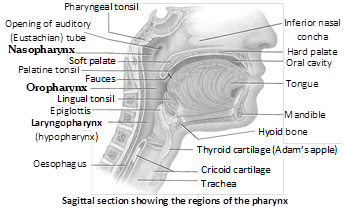
Nasopharynx lined by ciliated pseudostratified epithelia, oropharynx and laryngopharynx lined by non keratinized epithelium. Mouth serves as an alternate route for air when nasal chambers are blocked. Foramen by which pharynx opens into larynx called glottis. In general it remains open. During swallowing it is closed. It provides passage for air. Pharyns leads into the oesophagus through an aperture called gullet. In general condition it remains closed and opens at the time of swallowing. During swallowing epiglottis closes the glottis.
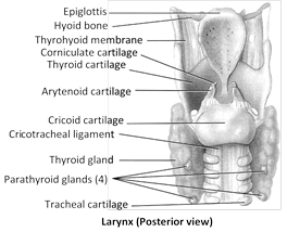
(iv) Larynx or Voice box : It is found both in frogs and rabbits. Larynx does not help in respiration. It is present on tip of trachea and is made up of 9 cartilages such as thyroid (single) has a prominence called pomum admi or adam’s apple, cricoid (single), arytenoid (paired), are piece of hyaline cartilage. While epiglottis (single), carniculate (paired), cuniform (paired), santorini are piece of elastic cartilage. Clinically, the cricoid cartilage is the landmark for making an emergency air way.
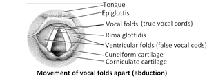
Larynx is a short tubular chamber and opens into the laryngopharynx by a slit like aperture called glottis. Glottis always remains open except during swallowing. Larynx is more prominent in men than women due to male hormone. Before puberty, the larynx is inconspicuous and similar in both sexes. Larynx is a voice producing instrument. For this purpose larynx have two types of vocal cord. In birds voice producing organ is syrinx, found at lower end of tracheae.
(a) False vocal cord or vibrating fold or anterior vocal cord : These are folds of mucus membrane. Gap between them is called rema vestibuli. These are not responsible for sound production. In elephants only true vocal cords are present and are responsible for this trumpet sound.
(b) True vocal cord or posterior vocal cords : They are made up of yellow elastic fibres. Gap between them is called rema glottides or peep hole. In males the length of true vocal cord is 2.25 cm and in female is 1.75 cm. Sound produced by rabbit is called quaking. Hippopotamus lacks true vocal cords. Pitch is controlled by the tension of vocal folds.

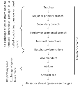
(v) Trachea : It is a tubular structure of about 12 cm. in length and 2.5 cm in diameter. The wall of trachea is made of fibres, cartilage muscles and the mucus membrane. In middle of thorax at the level of 4th and 5th thoracic vertebra it divides into two branches called right and left primary bronchi. Further division of primary bronchi is given in form of arrow diagram.
(a) Kultchitsky cells or argentaffin cells : They secrete serotonin and histamine. Histamine dilate while serotonin constrict the bronchioles.
(b) Clara cells : They secrete a phospholipid named diapalmityl lecithin which acts as a surfactant. This surfactant prevents the collapse of bronchioles lacking cartilagenous rings. Collapsing of lungs is called atelectesis. Pottle in 1956 proved the existence of surfactant. Surfactant is formed by clara cells only at later stage of foetal life. Some times at birth some infants are devoid of surfactant so there is great respiratory difficulty because lungs refuse to expand. In this condition death may occur. This is called respiratory distress syndrome (RDS) or hyaline membrane disease (HMD) or glassy lung disease.
(c) Dust cells : They are phagocytes which eat foreign particles (dust).
Different epithelium living in respiratory tract
| Vestibular region of nose | Skin having hair |
| Respiratory region of nose | Ciliated pseudostratified |
| Olfactory region of nose | Olfactory (Schneiderian) epithelium |
| Pharynx (Oropharynx, Laryngopharynx) | Non-keratinised stratified squamous |
| Trachea and bronchi (Upper) | Pseudostratified ciliated columnar epithelium with mucus cells |
| Lower bronchi (Secondary / Tertiary) | Lined by simple ciliated columnar epithelia |
| Terminal bronchioles and beginning of respiratory bronchiole | Simple ciliated columnar epithelium without mucus cells |
| Rest of respiratory bronchioles, alveolar duct | Non ciliated cuboidal epithelium |
| Alveoli | Non ciliated squamous |
| Alveoli of frog?s lungs | Columnar ciliated epithelium |


You need to login to perform this action.
You will be redirected in
3 sec

