Heart
Category : 11th Class
The form, structure and function of heart exhibits much variation. The characteristics of heart of fishes, amphibians, reptiles, birds and mammals is presented in the following table.
Heart of vertebrates
|
S.No. |
Class of vertebrates |
Characteristics |
Example |
Diagram |
|
1. |
Pisces (= Branchial heart), Cyclostomata |
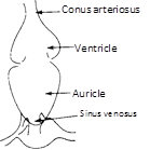
Thick, muscular, made of cardiac muscles, has two chambers (i) auricle and (ii) ventricle. The heart is called venous heart since it pumps deoxygenated blood to gills for oxygenation. This blood goes directly from gills to visceral organs (single circuit circulation). A sinus venosus and conus arteriosus is present. Lung fishes have only one auricles and one ventricle. |
Labeo Scoliodon
|
|
|
2. |
Amphibians, Lung fish |
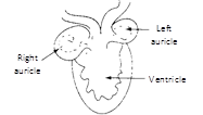
Heart consists of : (i) Two auricles (ii) Undivided ventricle (iii) Sinus venosus (iv) Truncus arteriosus (conus + proximal part of aorta) Right auricle receives blood from all the visceral organs (deoxygenated) via precaval and post caval. Pulmonary artery carries deoxygenated blood to lungs for oxygenation. This blood returns to left auricle via pulmonary vein (Double circuit circulation) (v) S.A. node in sinus venosus (vi) Trunchus arteriosus divided into synangium, pylangium |
Frog Toad Neoceratodus or Dipnoi |
|
|
3. |
|
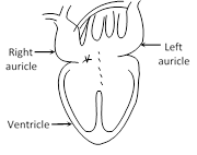
Heart consists of : (i) Left and right auricle (ii) Incompletely divided ventricle (Ventricle in crocodiles, gavialis, and alligator is completely divided) (iii) Sinus venosus (iv) Conus arteriosus divided into right systemic, left systemic and pulmonary arch. (Double circulation) (v) Foramen panizzae at crossing of right-left systemic arch. (vi) Only SA node in right auricle |
Lizards Snakes Turtles |
|
|
4. |
Aves |
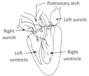
Exhibit double circulation : Heart consists of (i) Left and right auricle (ii) Left and right ventricle (iii) Complete separation of arterial and venous circulation (iv) Only right systemic arch is present (v) Sinus venosus and truncus, arteriosus absent (vi) Two pace maker SA node and AV node (vii) Mitral valve present. |
Pigeon
|
|
|
5. |
Mammals |
Same as bird except that mammals have left systemic arch. |
Rabbit, man |
|
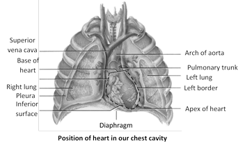
Shape and position : Reddish, roughly conical, highly muscular, mesodermal hollow organ of the size of one's fist. Its average weight in males is about 300 gm. and in females about 250 gm. It lies behind the sternum in the mediastinum space of thoracic cavity in between the two lungs. The broader base faces upward and backward. The narrower apex is directed downward, forward and slightly towards left, lying between 5th and 6th ribs and rests on the diaphragm. The heart is about 12 cm (5 inch) long, 9 cm (3.5 inch) wide and 6 cm (2.2 inch) thick.
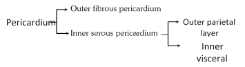
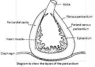
Protective covering : Heart is enclosed in a tough, 2 layered fibroserous sac, the pericardium. The outer layer is non-distensible fibrous pericardium and inner layer is thin serous pericardium which further consists of outer parietal layer (attached to fibrous pericardium) and inner visceral layer (adhered to the heart).
Between the parietal and visceral layers, occurs a narrow potential space, the pericardial cavity which is derived from coelom and is filled with serous pericardial fluid for frictionless movement and protection from shock and mechanical injury.
Histology : The heart wall consists of connective tissue, blood vessels and cardiac muscle fibres in 3 different layers - Epicardium, Myocardium and Endocardium.
(1) Epicardium : The outermost epicardium, also called visceral layer of the serous pericardium, is the thin, transparent outer layer of the wall. It is composed of mesothelium and connective tissue. Visceral pericardium, joined to myocardium by connective tissue.
(2) Myocardium : Middle, highly vascular layer, composed of cardiac muscle fibres joined together by intercalated disc. The connective tissue in myocardium acts as cardiac skeleton. Myocardium is thickest where the endocardium is thinnest.
(3) Endocardium : Innermost layer lining the cavity of heart and consisting of endothelium of squamous cells resting on thin basement membrane of loose connective tissue.
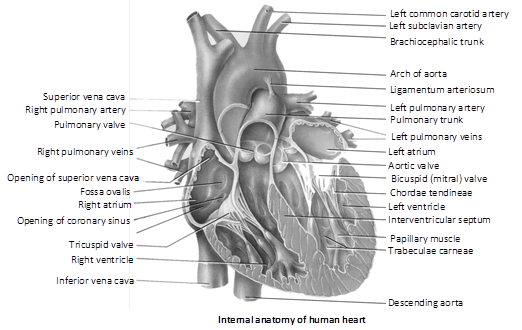
External structure : Human heart is 4-chambered and is divided by septa into two halves - right and left. Each half has one darker, thin walled auricle in the broader upper region and one lighter, thick-walled ventricle in the narrower lower region.
Sinus venosus and conus/truncus/bulbus arteriosus are accessory chambers in the heart of lower vertebrates (fishes and amphibians). In rabbit, sinus venosus is formed in the embryo but later it becomes a part of wall of right auricle.
In frog, sinus venosus spreads upon most of the dorsal side of heart and conus arteriosus lies obliquely upon the ventral surface of right atrium.
Internal structure
(1) Auricles : Atria are thin walled. They act as reservoirs for blood entering the heart. Right auricle is bigger than left auricle and both are separated by a myomembranous partition called Interatrial or interauricular septum. During embryonic stage, at the place of this septum, there are present septum primum and septum secondum having a gap (aperture) called foramen ovale between them. From the opening of inferior vena cava upto foramen ovale, there is a flap called Eustachian flap which prevents the blood in the foetal heart go to lungs because in foetal life, lungs are not functional purification of blood is done by placenta.
At the time of birth, there is closure of foramen ovale but there remains depression on posterior part of the right surface of interauricular septum in rabbit. In man this depression is present on both the side. because of least regenerative power in human being. The depression towards right atrium is called fossa ovalis and depression towards left atrium is called fossa lunata.
The inner surface of auricles is smooth. A network of muscular ridges called musculi pectinati or trabeculi pectinati occurs internally in the region of the auricular appendages and give comb like appearance.
• PFO (Patent Foramen Ovale) or septal defect : In case there is no closure of foramen ovale, then disease is called PFO. In this condition, there is mixing of blood after birth which gives bluish appearance to the body called as Cyanosis. Such child is called Blue Baby.
(2) Ventricles : The right and left ventricles are demarcated by an interventricular septum which is obliquely curved towards right, so that the left ventricle is larger than right one. However, the cavity of left ventricle is relatively smaller and nearly circular because the myocardium of left ventricle is 3 times thicker than right ventricle whose cavity is larger and somewhat crescentic.
The walls of the ventricles are internally raised into a number of thick, muscular, column shaped projections called columnae carnae or trabecular carnae; and a few large muscular elevations called papillary muscles or musculli papillares which are 3 in right ventricle and 2 in left ventricle. These muscles act as anchors for chordae tendinae.
Numerous, strong, inelastic thread like tendons present in the mammalian heart but absent in frog.
• Regurgitation : If there is weakening of papillary muscles or breaking of chordae tendinae, then AV valves revert into auricles. So, blood goes in opposite direction, it is called regurgitation. Sometimes, there is narrowing of valves. So, there remains gap between the valves which causes regurgitation.
? Moderator band : Right ventricle contains a prominent muscular trabeculum called moderator band which extends from the interventricular septum to anterior papillary muscle.
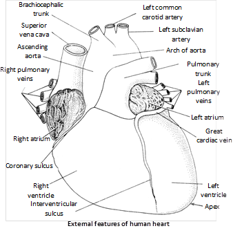
Major blood vessels associated with heart : The blood vessels that enter or leave the heart are called Great Blood Vessels.
(1) Superior vena cava or precaval : Brings deoxygenated blood from head and upper parts of the body into the right auricle through an opening which is single in human and cat and two in rabbit as there are 2 precavals - right and left in rabbit.
(2) Inferior vena cava or post caval : Drains deoxygenated blood from middle and lower parts of the body into the right auricle through a single opening which is bordered by a membranous, falciform fold which is a remnant of the foetal valve of Eustachian.
(3) Coronary sinus : Returns deoxygenated blood from heart wall into right auricle through a single opening.
(4) Pulmonary vein : Four pulmonary veins, two from each lung, carry oxygenated blood from the lungs and open into the left auricle through four openings. In rabbit, the pulmonary veins open in the left auricle through 2 openings.
(5) Pulmonary aorta/arch : Arises from upper left corner of right ventricle through a single opening and divides into right and left pulmonary arteries which carry deoxygenated blood to the lungs for oxygenation.
(6) Systemic aorta : Arises from upper right corner of left ventricle through a single opening and has 3 regions ? ascending aorta, arch of aorta and descending aorta. It distributes oxygenated blood to various body parts except lungs.
• Ligamentum arteriosus : During foetal life, because the lungs are non-functional hence blood of pulmonary aorta comes into systemic aorta through a small duct called ductus botalli or ductus arteriosus soon after birth, deposition of elastin fibre blocks this duct, forming a new structure called ligamentum botalli or ligamentum arteriosus.
• PDA (Patent Ductus Arteriosus) : If the ligamentum arteriosus remains open, the condition is called PDA. In this case, there is mixing of blood which leads to blue baby.
Valves : The valves present in the mammalian heart are tendinous cords.
(1) Eustachian valve : Present on the opening of inferior vena cava (post caval) in the right auricle in rabbit, whereas in human, the vestige of eustachian valve is present over the opening of post caval vein. It allows the passage of blood in right auricle.
(2) Haversian valve : Present in human but absent in rabbit. It is present over the opening of precaval vein and allows the passage of blood in right auricle.
(3) Thebesian or coronary valve : Present over the opening of coronary sinus in right auricle in mammals and allows the passage of blood in right auricle.
(4) Right A.V. valve or Tricuspid valve : Present between right auricle and right ventricle. It consists of 3 membranous flaps or cusps.
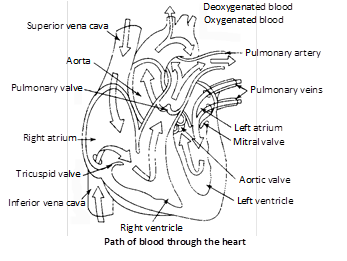
(5) Left A.V. valve or Bicuspid or Mitral valve : Present between left auricle and left ventricle. It consists of 2 flaps or cusps. The bicuspid valve resembles mitre or topi of bishop, hence, also called as Mitral valve.
(6) Semilunar valves : At the base of pulmonary arch and systemic aorta, three membranous, pocket-shaped flaps called semilunar valves are present. They allow the passage of blood from ventricles to respective blood vessels, but prevent the return of blood.
Nodal tissue : The nodal tissue consists of the following ?
(1) Sinu-auricular or S.A. node : Also called as pacemaker, node of keith and flack, heart of heart, brain of heart, pulsation centre. It is located in the right wall of right atrium below the opening of superior vena cava. This is the place where sinus venosus is incorporated in the wall of right atrium in the embryo. S.A. node is the main tissue of heart and has highest degree of autorhythmicity (generates beating impulse at the rate of 70-80 times/minute) but least conductivity. The rhythmic impulses produced are called as Sinus rhythmia. In frog S.A. node is present in sinus venosus.
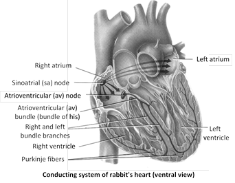
(2) Atrio-ventricular node or A.V. node : Also called reserve pacemaker, node of Twara and Aschoff. Discovered by Lewis Kent. It lies in the right atrium near the junction of interauricular and interventricular septum close to the opening of coronary sinus. It is concerned with the conduction of cardiac impulses generated by S.A. node, but it can also generate the impulse at the rate of 40-60/minute. These impulses produced are rhythmic and called nodal rhythmia. In frog, A.V. node is absent.
(3) Bundle of His or A.V. bundle : Discovered by His. It arises from A.V. node, descends in the interventricular septum and bifurcates into two branches innervating the wall of right and left ventricle respectively. The myocardium of atria and ventricles are discontinuous and this bundle is the only muscular connection between the two. It is concerned with the conduction of impulse from atria to the tip of ventricle but can also generate impulse at the rate of 35-40/minute. The impulses produced are non-rhythmic.
(4) Purkinje fibres : Numerous, modified muscle fibres which act as sympathetic nerve fibres. They arise from branches of bundle of His and provide impulse to myocardium of ventricles. They can also generate non-rhythmic impulse at a rate of 30-35/minute.
Working of nodal tissue : S.A. node spontaneously initiates a wave of contraction which is conducted along the tracts of special muscle fibres called internal pathways over both the auricles at a rate of 1m/sec. The impulse generated travels first in the right atrium than in left atrium. So, right atrium contracts first but the contraction ends simultaneously in both atria. As the musculatures of atria and ventricles are discontinuous and are separated by a septum of fibrous connective tissue, called annular pad in mammals, the wave of contraction is received by A.V. node from myocardium of atria and is provided to bundle of His. The impulses reach the A.V. node about 0.03 seconds after their origin from S.A. node. The A.V. node generates a fresh wave of contraction which passes over both the ventricles along the bundle of His and its ramifications at the rate of 1.5 to 4 m/sec. The Purkinje fibres bring about the contraction of ventricles from the apex of heart which passes quickly towards the origin of pulmonary and systemic arches forcing blood into them.
S.A. node not only acts as pacemaker but also establishes the basic rhythm at which the heart beats. In case of degeneration of S.A. node, A.V. node can generate impulse but it will lead to abnormal beating (arrhythmia). The failure of atrial impulse to pass into ventricles for a few seconds to few hours is called ventricular escape or stokes-adams syndrome leading to delayed pick up of heart beat. In such conditions, artificial pacemaker (Lithium Battery) is placed underneath the patient?s chest.
• Ectopic pacemaker : If any cardiac muscle other than the conducting tissue (nodes) generates impulse, then extra beats are heard. Such muscles are called Ectopic pacemaker.
In mammals, conducting system of the heart has S.A. node, A.V node and complicated system of conducting fibres. But in frog, it has only S.A. node and system of conducting fibres is simple.
Heart beat : The spontaneous and rhythmic contraction and relaxation of the heart to pump out and receive blood to and from the body is called Heart beat. Depending upon the nature of control of the heart beat, hearts are of 2 types - Neurogenic and Myogenic or autorhythmic.
Differences between Neurogenic heart and Myogenic heart
|
S. No. |
Neurogenic heart |
Myogenic heart |
|
1. |
The heart beat is initiated by a ganglion situated near the heart. |
The heart beat is initiated by a patch of modified heart muscle. |
|
2. |
The impulse of contraction originates from nervous system. |
The impulse of contraction originates itself in the heart. |
|
3. |
The heart normally stops beating immediately after removal from the body. Therefore, heart transplantation is not possible. |
The heart removed from the body continues to beat for some time. Therefore, heart transplantation is possible. |
|
4. |
Examples : Hearts of some annelids and most arthropods. |
Examples : Hearts of molluscs and vertebrates. |
Origin and conduction of heart beat : Initiation of heart beat is under special bundles of cardiac muscles called nodal tissue or autorhythmic cells. They act as pace maker so setting the rhythm for the entire heart and they form conducting system.
Heart beat rate : Heart beat/minute or number of cardiac cycles/minute. Females have higher heart rate than males.
Normal heart beat rate\[\to \]Rhythmia
Abnormal heart rate\[\to \]Arrhythmia
Decrease in heart rate\[\to \]Bradycardia
Increase in heart rate\[\to \]Tachycardia
Heart beat rate in some animals
Elephant - 29/min.
Human - 70-80/min.
Foetus (human) - 140-150 / min.
New born baby - 115-130/min.
Horse - 35-40/min.
Rat - 300-500/min.
Frog - 64/min.
Rabbit - 200/min.
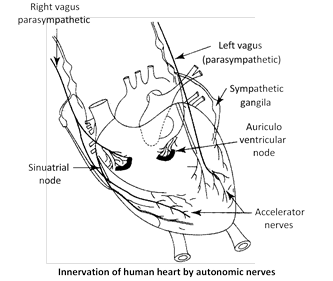
Regulation of heart beat : The centre controlling the heart rate (cardiac centre) is present in medulla oblongata of brain and possess chemoreceptors sensitive for \[C{{O}_{2}},{{O}_{2}}\] and also for blood pressure. This centre is under the influence of hypothalamus which is the controller of autonomic activities.
(1) Nervous control : Brain receives two sets of nerve fibres : Sympathetic and para sympathetic or vagal.
When there is increase in blood \[C{{O}_{2}},\] the sympathetic nerve fibres stimulate S.A. node by producing sympathin (adrenaline + noradrenaline). This compound induces impulse generation by inducing entry of \[C{{a}^{2+}}\]into cardiac muscles. So, heart beat and force of contraction increase (Tachycardia). After action, sympathin is destroyed by sympathenase, COMT (catechol orthomethyl transferase) and MAO (Mono Amino Oxidase).
When there is increase in blood \[{{O}_{2}},\] the parasympathetic or vagal (10th cranial) nerve inhibits S.A. node by producing acetylcholine. This compound increases contraction time and hence, heart beat is decreased (Bradycardia). After action, acetyl choline is destroyed by enzyme acetyl choline esterase (AchE). This chemical regulation of heart beat on behalf of nerves was discovered by Otto Loewi.
• Vagus escape : Stimulation of vagus nerve decreases the heart rate but its continuous stimulation shows no further decrease. This phenomenon is called Vagus escape.
(2) Hormonal control : Hormones from adrenal medulla adrenaline and nor adrenaline accelerate the heart beat, the latter under normal conditions and the former at the time of emergency. Thyroxine hormone also increases the heart beat by increasing energy production.
• Pounding : Very fast heart beat during some conditions like anger and love.
Factors affecting heart rate
(1) Heart rate increases with increase in basal metabolic rate (BMR).
(2) Heart beat rate increases as the size of the animals body decreases.
(3) Decrease in pH also increases heart rate.
(4) Heart rate increases with increase in temperature.
(5) Increase in \[N{{a}^{+}}\] ions in blood or in cardiac muscles, decrease heart rate.
(6) Increase in \[C{{a}^{2+}}\] ions in blood increase heart beat but if they are injected in cardiac muscles, heart stops in contracted phase which is called Systolic Arrest.
(7) Injection of \[{{K}^{+}}\] ions in heart muscles stop impulse generation. So, heart stops in diastolic or Relax phase.
(8) \[{{H}^{+}}\]ions reduce force of contraction of heart.
(9) Increased inspiration, muscular exercise, low oxygen tension, injection of adrenaline, thyroxine, sympathin - all increase heart rate.
(10) Increased expiration, during sleep, injection of acetylcholine decrease heart rate.
(11) Stenosis - Narrowing of valve is called stenosis.
(12) Alkalosis - Decreases heart rate.
(13) Anoxia - (Absence of \[{{O}_{2}}\] in tissue) Increases heart rates.
(14) \[C{{O}_{2}}\] more amount, decreases heart rate.
(15) \[C{{O}_{2}}\]moderate amount, increases heart rate.
(16) Epinephrine and nor epinephrine increase heart rates.
(17) Thyroid hormone increases heart rate.


You need to login to perform this action.
You will be redirected in
3 sec