Digestive System of Human
Category : 11th Class
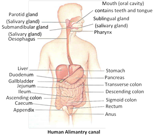
Digestion in vertebrates occurs in the digestive tract or alimentary canal. The various parts involved in digestion can be broadly grouped in two groups –
(1) Digestive tract or alimentary canal
(2) Digestive glands
(3) Digestive tract or alimentary canal
On the basis of the embryonic origin, the alimentary canal of vertebrates can be divided into three parts –
(1) Fore gut / Stomodaeum : Ectodermal. It includes buccal cavity / oral cavity, pharynx, oesophagus, stomach and small part of duodenum.
(2) Mid gut / Mesodaeum : Endodermal. It includes small intestine, and large intestine.
(3) Hind gut / Proctodaeum : Ectodermal. It includes anal canal and anus.
Parts of alimentary canal and its histology
Mouth
The mouth is a transverse slit bounded by two movable lips or labia, upper lip and lower lip. Upper lip has small ridges on the sides, a tubercle in the middle and a vertical groove (philtrum) above.
It is a narrow space between lips and gums in front and gums and cheeks on the sides. Its lining contains mucous glands. In the vestibule, a small median fold of mucous membrane, the superior labial frenulum, connects the middle of the upper lip to the gum and usually a similar but sma0ller inferior labial frenulum connects the middle of the lower lip to the gum.
It includes anterior buccal cavity lined by stratified squamous epithelial cell and posterior pharyngeal cavity lined by columnar epithelial cell. It is distinguished into three region. Pharynx is a vertical canal beyond the soft palate. The food and air passages cross here. Pharynx may be divided into three parts; Nasopharynx, Oropharynx and Laryngopharynx.
Main structures of Buccopharyngeal cavity are –
(1) Fauces : A triangular area present between buccal cavity and pharynx in human.
(2) Palate : The roof of buccal cavity is called Palate. In crocodiles and mammals horizontal shelf like processes of premaxilla and maxilla and the palatine bones of upper jaw fused and form a secondary palate. Which separates the buccal cavity from nasal cavity. Palate is distinguished into three regions –
(i) Hard palate : Anterior, bony portion formed of maxilla and palatine bones in human and premaxilla, maxilla and palatine bones in rabbit. Hard palate have transverse ridges called palatine rugae. Such rugae or ridges are more develop in carnivorous mammals because their function is to firmly grip the food and prevent it from slipping out the cavity.
(ii) Soft palate : Posterior soft part, made up of connective tissue and muscles.
(iii) Vellum palati/uvula : Posterior most part of soft palate, which hangs in the region of pharynx. It closes the internal nostrils during degglutition.
(3) Palatine glands : Numerous mucous glands. Chiefly present in soft palate, secretes mucous for lubrication.
(4) Naso-palatine duct : One pair, present in rabbit, extends from nasal passage to the buccal passage, contains Jacobson’s organ concerned with olfaction.
(5) Vibrissae : A tuft of hairs on upper lip of rabbit.
(6) Hare-cleft : A cleft on the upper lip of rabbit, which makes it bilobed.
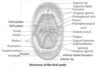
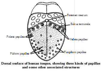
(7) Tongue (linguae) : Ectodermal, single, pinkish, oval, elongated highly muscular (mesodermal) and protrusible present on the floor of buccopharyngeal cavity the cells present are stratified squamous epithelial cells. A furrow termed the sulcus terminalis divides the oral part and pharyngeal part of the tongue. The limbs of the sulcus terminalis run laterally and forward from a median pit, named the foramen caecum.
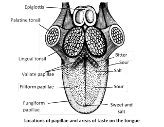
Posterior part of tongue (endodermal) is attached with hyoid, middle one with the floor of buccopharyngeal cavity with the help of frenulum lingum and anterior part is free. The tongue is provided with two specialized structure viz. lingual papillae and lingual glands or weber’s gland. Lingual glands are the mucous glands, which secretes mucous. Lingual papillae are numerous, minute projections chiefly present on the dorsum of the tongue. All these lingual papillae can be grouped as simple lingual papillae and taste papillae. Taste papillae are of following types –
(i) Circumvallate : Circular largest in number, present in the posterior part of the tongue extending from one side to another. They possess taste buds. These are the largest of all the papillae.
(ii) Fungiform : Mushroom shaped (Fungi - shaped), numerous, present at the anterior margins and tip of the tongue. They have 200 taste buds.
(iii) Foliate : Leaf like flat, less in number, present at the posterior margin of the tongue. They are absent in human and found in rabbit.
(iv) Filiform : Conical shaped, smallest and most numerous distributed throughout tongue. They are without taste buds.
Hence, in human taste is recognized with the help of circumvallate and fungiform taste papillae. In man the anterior end of tongue feels sweet taste, posterior part feel bitter taste, sides feel sour taste and a small part behind the anterior end feel salty taste.
Functions of tongue : Important function of tongue are as follows –
(i) Acts as universal toothbrush, as it helps in tooth cleaning.
(ii) Helps in speaking.
(iii) Helps in degglutition.
(iv) Helps in mixing saliva with food.
(v) Acts as a curry comb in many animals, hence help in body cleaning.
(vi) Helps in taste detection.
(vii) In dog helps in regulation of body temperature. The phenomenon is called as “Panting”.
(viii) In frog and other animals, it helps in prey capturing
(8) Teeth : Teeth is a living structure. On the basis of embryonic origin, teeth in vertebrates are of following two types-
(i) Horny/ectodermal/epidermal/false teeth : The teeth which develops only from ectoderm. Examples -
Cyclostomes, tadpole larva of frog, prototherian mammals etc.
(ii) True teeth : The teeth which develops from both ectoderm and mesoderm. Examples – Fishes, amphibians, reptiles, eutherian mammals etc.
Differentiation of teeth : Morphologically, teeth can be distinguished as homodont or heterodont.
(i) Homodont : When all the teeth are structurally and functionally similar. Examples – Vertebrates except metatherian and eutherian mammals.
(ii) Heterodont : When the teeth are different in structure and functions. They are distinguished into four types incisors, canines, premolars and molars. Examples – metatherian and eutherian mammals.
(a) Incisors : These are the front teeth borne by the premaxillae in upper jaw and tips of dentaries in lower jaw. They are single-rooted monocuspid and long, curved and sharp-edged. They are adapted for cutting or cropping and biting.
(b) Canines : There is one pointed canine in each maxillary of upper jaw and each dentary of lower jaw next to the incisors. They are meant for piercing, tearing and offence and defence. They are single rooted and monocuspid.
(c) Premolars : They have one root (only in upper first PM two roots) and two cusps (bicuspid). They are meant for crushing, grinding and chewing.
(d) Molars : They have more than two roots (upper molars have three roots and lower molars have two roots) and 4 cuspid.
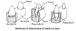
Attachment of teeth : On the basis of attachment of teeth at their bases with the jaw bones, teeth can be differentiated into –
(i) Acrodont : Teeth are attached to the free surface or summit of the jaw bone, as in a shark or frog. Such teeth are apt to break off easily but are replaced.
(ii) Pleurodont : In this condition, common in urodeles and lizards, teeth are attached to the inner side of jaw bone by their base as well as one side.
(iii) Thecodont : Such teeth are characteristic of mammals. Teeth have well developed roots implanted in deep individual pits or socketes called alveoli or theca, in the jaw bone. These type of teeth also present in crocodilians, fossil toothed bird (Archeaeopteryx).
Succession of teeth : According to their replacement (succession), teeth can be divided into 3 categories: polyphyodont, diphyodont and monophyodont.
(i) Polyphyodont : In lower vertebrates, teeth can be replaced an indefinite number of times during life. e.g., – Fishes, amphibia, reptilia.
(ii) Diphyodont : In most mammals teeth develop during life in two successive sets, a condition known as diphyodont. Teeth of the first set are known as deciduous teeth or milk teeth or lacteal teeth whereas the second set is called permanent teeth.
(iii) Monophyodont : In some mammals such as platypus, marsupials, moles, sirenians, toothed whale etc. only one set of teeth develops known as monophyodont condition.
Types of cheek teeth
(i) Bunodont : Crown with small, blunt and round cusps as in man, monkey, pig etc. found in mixed diet mammals.
(ii) Secodont : With sharp cutting edges for tearing flesh as in carnivores.
(iii) Lophodont : Only one cusp is present with transverse ridges called lophos, e.g., Elephant.
(iv) Selenodont : With vertical crescentic cusps as in grazing mammals like cow, sheep and goat. Selenodont teeth are two types –
(a) Brachyodont : Normal low crowned selenodont teeth with large roots are termed brachyodont. e.g. Ground squirrel, cattle.
(b) Hypsodont : In large grazing mammals teeth are elongated, prism shaped with high crown and low roots. e.g. Horse.
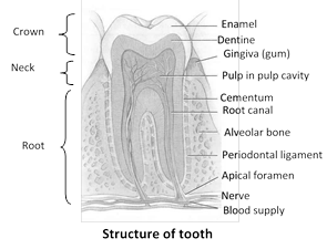
Structure of teeth : Teeth divided into three parts –
(i) Root : Inner most, attached to the bone with help of cement (hyaluronic acid).
(ii) Neck : Middle, small, covered with gum. Gum provides strength to the teeth.
(iii) Apex or crown : External exposed part of teeth. Longest part, white in colour.
A small cavity present inside teeth called as pulp cavity or dentine pulp cavity. It contains blood vessels, lymphatic vessels, nerve fibres, connective tissue etc. and provides nutrition to odontoblast cells or osteoblast cells. The odontoblast cells are mesodermal in embryonic origin forming immediate covering of the pulp cavity. The cells secrete dentine/ivory. Bulk of tooth in a mammal is formed of dentine. Dentine is a layer of inorganic substances \[(62-69%),\] which surrounds the odontoblast cells. It is mesodermal in origin. Enamel, secreted by Ameloblast/Enameloblast cells, forms the outermost covering. It is ectodermal and made up of 92% of inorganic substances, hence considered as hardest part of the body. The inorganic substances present are \[[C{{a}_{3}}{{(P{{O}_{4}})}_{2}},Ca{{(OH)}_{2}}.\,\,{{H}_{2}}O]\] Calcium phosphate (85%), Calcium hydroxide and Calcium Carbonate. Cement/Cementum attaches the tooth root to the bone.
Dental formula : Each mammalian species is characterized by its own specific dentition with a definite number and arrangement of teeth. Hence, dentition is of taxonomic importance. It is expressed by a dental formula as below –
Rabbit : \[i\frac{2}{1},c\frac{0}{0},\,pm\frac{3}{2},m\frac{3}{3}=\frac{8}{6}\times 2=28\] or briefly,
\[\frac{2033}{1023}=\frac{2+0+3+3}{1+0+2+3}\times \frac{2}{2}=\frac{16}{12}=28\]
(i = incisors; c = canines; pm = premolars; m = molars)
Dental formulae of some common mammals
|
Horse and pig |
\[\frac{3.1.4.3}{3.1.4.3}\times \,2\,=44\] |
Cat |
\[\frac{3.1.3.1}{3.1.2.1}\times \,2\,=30\] |
|
Dog |
\[\frac{3.1.4.2}{3.1.4.3}\times \,2\,=42\] |
Squirrel |
\[\frac{1.0.2.3}{1.0.1.3}\times \,2\,=22\] |
|
Lemur |
\[\frac{2.1.3.3}{2.1.3.3}\times \,2\,=36\] |
Rat |
\[\frac{1.0.0.3}{1.0.0.3}\times \,2\,=16\]. |
|
Man (adult set) |
\[\frac{2.1.2.3}{2.1.2.3}\times \,2\,=32\] |
Elephant |
\[\frac{1.0.0.3}{0.0.0.3}\times \,2\,=14\] |
|
Cow |
\[\frac{0.0.3.3}{3.1.3.3}\times \,2\,=32\] |
Human set (milk set) |
\[\frac{2.1.0.2}{2.1.0.2}\times \,2\,=20\] |
Oesophagus (food tube)
(1) Morphology : Single, ectodermal, dorsal to trachea, approximately 25 cm long. passes through thoracic cavity and opens into stomach present in abdominal cavity. Oesophagus anteriorly opens into pharynx through gullet and posteriorly into stomach through cardiac orifice.
(2) Histology : Serosa is absent but outermost layer of connective tissue is called as tunica adventitia. Muscular layer are striated/voluntary in anterior region and unstriated/involuntary in posterior part. The epithelial lining is made up of non-keratinized stratified squamous epithelial cells. Goblet cells are present.
Function : Conduction of food.
Stomach
(1) Structure : Single oval, elongated, unilobed present within abdominal cavity below diaphragm. It consists of three parts as cardiac/fundic (anterior), corpus/body (middle, chief part) and pyloric (posterior part) in human, whereas in rabbit stomach is bilobed and consists of three parts as cardiac (Anterior), fundic (middle, chief part) and pylorus (posterior). Two types of valves are present in the stomach viz. Cardiac sphincter valve between oesophagus and stomach and pyloric sphincter valve between stomach and duodenum. It new born baby cardiac sphynctor is much less developed that is why regurgitation of gastric contents is very common. Inner surface of stomach raised into numerous longitudinal folds called gastric rugae. In case of ruminant mammals (cud chewing mammals) oesophagus consists of only skeletal or voluntary muscles.
Oesophagus lack digestive glands but multicellular glands are found, which extends upto submucosa. Due to the presence of these submucosal mucous glands, submucosa of oesophagus is thickest than other parts of alimentary canal.
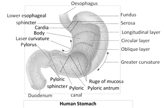
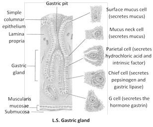
(2) Histology : Outermost layer is serosa. Muscular layer is three layered with outer longitudinal, middle circular and inner oblique. Muscles are involuntary and unstriated. Epithelial lining is made up of simple columnar epithelial cells and specialized cells present in the gastric glands. The nomenclature of gastric glands is according to the parts of the stomach. The various type of gastric glands and the cells present in them are as follows –
(i) Anterior part : Cardiac gastric glands in rabbit and human cells present are mucous neck cells secreting mucous.
(ii) Middle part : Fundic gastric/Main gastric glands glands in rabbit and corpus in human has at least four distinct types of cells –
(a) Peptic or zymogenic or chief or central cells : Secretes two digestive proenzymes pepsinogen and prorennin.
(b) Oxyntic or parietal cells : Secretes \[HCl\] and castle's intrinsic factor required for the absorption of vitamin B12. Hyperacidity is a abnormally high a degree of acidity due to the secretion of large quantity of \[HCl\] i.e. gastric juice.
(c) Mucous neck cells : Secretes alkaline mucous.
(d) Argentaffin cells or Kultchitsky or enterochromaffin cells : Responsible for the secretion of vasoconstrictor seratonin.
(iii) Posterior part : Pyloric gastric glands in rabbit and human-cells are mucous neck cells secreting mucous and some cells, called “gastrin” or “G” cells, secrete a hormone, named gastrin, which increases the motility of gastric wall and stimulates gastric glands for active secretion.
Functions
(1) Storage of food.
(2) Trituration or churning of food to mix with gastric juice.
(3) Functions of gastric juice (discussed along with gastric juice).
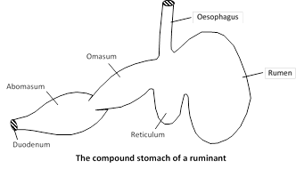
Stomach of ruminants (cud-chewing mammals) : The stomach of cattles have four parts, as rumen (paunch), reticulum(honeycomb), omasum (psalterium) and abomasum (rennet). Some authors believe that first three chambers are parts of the oesophagus, the fourth chamber is the real stomach secretes \[HCl\] and enzymes. The embryological studies have proved that all the chambers are parts of the real stomach. Camel and deer lack omasum. Reticulum is the smallest part and its cells are provided with water pockets for the storage of metabolic water.
In the rumen, food undergoes mechanical and chemical breakdown. Mechanical breakdown results from through churning brought about by muscular contractions and aided by cornified surface of villi. Chemical breakdown is caused by symbiotic microorganisms (bacteria and ciliates) that release enzyme cellulase, which act on cellulose and simplify it into short-chain fatty acids, such as acetic acid, butyric acid, propionic acid. This is called microbial digestion.
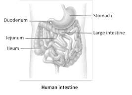
Small intestine
(1) Structure : Endodermal, longest part of alimentary canal present in the abdominal cavity, supported by a peritoneal membrane called mesentery. Wall of jejunum and ileum has circular or spiral internal fold called fold of kerckring or valvulae conniventes. Also numerous finger like projection called villi project from the wall of lumen, increasing internal surface area about ten time. The distal end of ileum is leads into the large intestine by ileo-caecal valve in man but in rabbit sacculus rotundus and ileo-coecal valve both are present.
(2) Parts : It is approximately 3 metres in human. It is divisible into three parts. In man small intestine divided into three parts :
Parts of small intestine
|
Duodenum (Proximal part) |
Jejunum (Middle part) |
Ileum (Posterior part) |
|
25 cm. Long Forming U-shaped loop before leading to jejunum, pancreas lies in the loop. |
About 1 m long and about 4 cm. wide. Wall is thicker and more vascular. Villi thicker and tongue-like. Plicae best developed. Peyer's patches are lacking. |
About 2 m long and about 3.5 cm. wide. Wall is thinner and less vascular. Villi thinner and finger-like. Plicae less developed. Peyer?s patches are present. |
(3) Histology : Serosa is the outer most covering. Muscular layer is generalized with involuntary, unstriated muscles. The cells present in the epithelial lining are simple columnar epithelial cells, which are brush- boardered i.e. provided with villi and microvilli to increase the surface area. The folds present are longitudinal and are called folds of kerckring or valvulae canniventes. Goblet cells secrete mucous. Payer’s patches are the oval, rounded masses of lymphatic tissue present in between lamina propria and epithelial lining. They produce lymphocytes. Brunner’s glands or Duodenal glands are the multicellular mucous glands present in the submucosa of duodenum only. They secrete mucous. In addition there are also found granular arogyrophil cell.
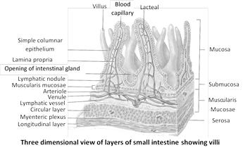
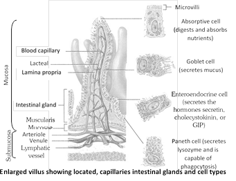
(4) Glands of small intestine : Various glands found in small intestine. Each gland has three types of cells : (1) Undifferentiated epithelial cell (2) Zymogenic cell (paneth cell) and (3) Argentaffin (Enterochromaffin cell).
Glands of small intestine
|
Brunner's glands |
Payer's patches |
Crypts of Leiberkuhn |
|
Found in duodenum only. Mucus secreating gland as so known as mucus gland. |
These are lymph nodules. They produce lymphocytes. Lymphocytes are phagocyte in nature which destroy harmful bacteria. |
Known as intestinal gland. Found in duodenum and ileum only. Secrete succus entericus i.e. intestinal juice. Formed by folding of lamina propia. |
Function : Digestion and absorption of food.
Large intestine
The name large intestine is due to large diameter (4-6 cm).
(1) Structure : Endodermal, approximately \[1.5-1.75\] metre long.
(2) Parts : They are following –
(i) Caecum : Spirally coiled 6 cm long in human and 45 cm long in rabbit. Its posterior end is present as a blind sac in abdominal cavity called vermiform appendix. Vermiform appendix is vestigeal but contains lymphatic tissue. Caecum in human is concerned with passage of food whereas in rabbit it is concerned with cellulose digestion and conduction of food.
(ii) Colon : Single endodermal approximately 1.3 m long in human distinguished into four limbs as ascending, transverse, descending and pelvic or sigmoid limb. Colon posses two specialized structures as Taeniae coli (present in the middle of colon) and Haustra, (dilated sac-like or pockets like structures surrounding taeniae). Colon is concerned with absorption of water of undigested food, 5%, salts, vitamins etc. hence concerned with faeces formation. Colon bacteria also synthesized vit. \[{{B}_{12}}\] and K.

(iii) Rectum : Single small dilated sac like in human whereas large beaded in rabbit. It is concerned with storage of faeces. Rectum has strong sphincter muscle in its wall. The sphincter keeps the canal as well as anus, closed when not used for defecation.
(iv) Function : Absorption of water from undigested food.
Anal canal and anus : Anal canal connects rectum with anus and it is about 3 cm. long. Anus is the terminal inferior opening of alimentary canal, which is guarded by an internal involuntary sphincter and an external voluntary sphincter.

The various types of digestive glands present in mammals are salivary glands, gastric glands, intestinal glands, pancreas and liver. The digestive glands secrete digestive juices. Parasympathetic nervous system increases the secretion of digestive juice whereas sympathetic nervous system decreases it.
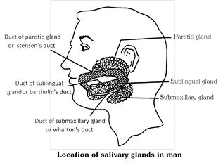
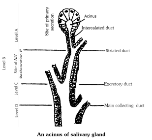
(a) Salivary glands : The three pairs of salivary glands present in humans are as follows –
(1) Parotid : One-pair, largest salivary gland present below pinna. A stenson’s duct arises from each gland, opening in vestibule between the 2nd molar teeth of upper jaw and checks. Parotid glands secrete enzymes. Viral infection of parotid glands causes “Mumps” (by paramyxo virus).
(2) Sub-mandibular / sub-maxillary : One-pair, present at the junction of upper and lower jaw in cheek region. A wharton’s duct arises from each gland and opens on lower jaw. These are seromucous glands.
(3) Sub-lingual : One-pair, present in the floor of buccopharyngeal cavity. These are mucous glands 6-8 ducts, called ducts of rivinus or Bartholin's duct arises from these glands and opens below tongue on the floor of buccopharyngeal cavity.
Saliva / salivary juice : The secretion of salivary glands is called saliva or salivary juice. Some of the characteristics are as follows –
(1) Amount : 1.0-1.5 litre/day
(2) Chemical nature : Slightly acidic.
(3) \[pH:\text{ }6.36.8\]
(4) Control of secretion : Autonomic reflex (parasympathetic nervous system increases salivation while sympathetic nervous system inhibit secretion.)
(5) Chemical composition : Water (99.5%), mucous (acts as lubricant), salts (\[NaCl,\text{ }NaHC{{O}_{3}}\]etc.), enzymes (ptyalin, lysozyme) etc.
Functions : Salivary juice and its enzymes –
(1) Makes the medium slightly acidic for the action of its enzyme.
(2) Help in taste detection, deglutition, speaking etc.
(3) Starch \[\underset{\text{(Salivary}\,\text{amylase)}}{\mathop{\xrightarrow{\text{Ptyalin/Diastase}}}}\,\] Maltose + Isomaltose + Limit dextrin.
(4) Bacteria (living) \[\xrightarrow{\text{Lysozyme}}\]Bacteria killed.
Gastric glands : There are approximately 35 million of gastric glands present in human stomach and grouped into three categories as already described along with stomach. The gastric gland secretes gastric juice.
Gastric juice
(1) Amount : 2-3 liters/day.
(2) Chemical nature : Highly acidic
(3) \[pH:\text{ }1.03.5\](due to presence of \[HCl\])
(4) Control of secretion : By gastrin hormone.
(5) Chemical composition : Water (99%), mucous, inorganic salts, castle’s intrinsic factor, \[HCl\] ( 0.5%, conc.) and enzymes prorennin and pepsinogen and gastrin lipase.
Functions of gastric juice and its enzymes
(1) Inactivates the action of ptyalin.
(2) Makes the medium acidic for the action of gastric enzymes.
(3) \[HCl\] kills micro organisms.
(4) \[HCl\] kills the living organism (prey etc.) if ingested.
(5) Pepsinogen (inactive) \[\xrightarrow{HCl}\] Pepsin (active).
(6) Prorennin (inactive) \[\xrightarrow{HCl}\] Rennin (active).
(7) Proteins + Peptones \[\underset{pH-1-3}{\mathop{\xrightarrow{\text{Pepsin}}}}\,\] Polypeptides + Oligopeptides.
(8)\[\underset{(\text{milk protein)}}{\mathop{\text{Casein}}}\,\underset{\text{C}{{\text{a}}^{\text{2}+}}}{\mathop{\xrightarrow{\text{Rennin}}}}\,\] Paracasienate Above phenomenon is called “curdling of milk”.
(9) Lipids \[\underset{negligible\,in\,human\,stomach\,acts\,at\,pH\,4-6}{\mathop{\xrightarrow{\text{Gastric}\,\text{Lipase}}}}\,\] Triglycerides + Monoglycerides.
(10) \[HCl\]is antiseptic.
(11) It act as preservative.
Lactose intolerance : Among mammals, man alone takes milk even after becoming adult. In some humans, secretion of lactase decreases or ceases with age. This condition is called lactose intolerance. Lactose intolerant persons fail to digest lactose of milk. In their large intestine, lactose fermented by bacteria, producing gases and acids.
Intestinal glands : Intestinal glands in mammals is a collective name for crypts of Lieberkuhn (secretes alkaline enzymatic juice) and Brunner’s glands (secretes mucous). Intestinal glands secrete intestinal juice.
Succus entericus (intestinal juice)
(1) Amount : 1.5 – 2.0 l/day.
(2) Chemical nature : Alkaline.
(3) \[pH:\text{ }7.6-8.3\]
(4) Control of secretion : Nervous and hormonal (Enterocrinin Duocrinin etc.)
(5) Chemical composition : Water (99%), mucous, inorganic salts, enzymes etc.
Function of Intestinal juice and its enzymes.
(1) Inhibits the action of gastric enzymes.
(2) Makes the medium alkaline for the action of it’s enzymes.
(3) Starch \[\xrightarrow{\text{Amylase}}\] Maltose + Isomaltose + limit dextrin.
(4) Maltose \[\underset{\left( \alpha \text{-glucosidase} \right)}{\mathop{\xrightarrow{\text{Maltase}}}}\,\] Glucose + Glucose.
(5) Isomaltose \[\xrightarrow{\text{Isomaltase}}\] Glucose + Glucose.
(6) Lactose (milk sugar) \[\underset{\left( \beta \text{-galactosidase} \right)}{\mathop{\xrightarrow{\text{Lactase}}}}\,\] Glucose +Galactose.
(7) Sucrose (cane sugar) \[\underset{\left( \beta \text{-fructosidase} \right)}{\mathop{\xrightarrow{\text{Sucrase / Invertase}}}}\,\] Glucose + Fructose.
(8) Polypeptides + Oligopeptides \[\underset{\left( \text{Amino}-\text{peptidase} \right)}{\mathop{\xrightarrow{\text{Erepsin}}}}\,\] Amino acids.
(9) Trypsinogen (inactive) \[\xrightarrow{\text{Enterokinase}}\] Trypsin (active).
(10) Lipids \[\xrightarrow{\text{Lipase}}\] Fatty acids + Glycerol + Monoglycerides.
(11) Phospholipids \[\xrightarrow{\text{Phospholipase}}\] phosphorous + Fatty acids + Glycerol + Monoglycerides.
(12) Organic phosphate \[\xrightarrow{\text{Phosphatase}}\] Free phosphate.
(13) Nucleic acid \[\xrightarrow{\text{Polynucleotidase}}\] Nucleotides.
(14) Nucleosides \[\xrightarrow{\text{Nucleosidase}}\] Nitrogenous bases.
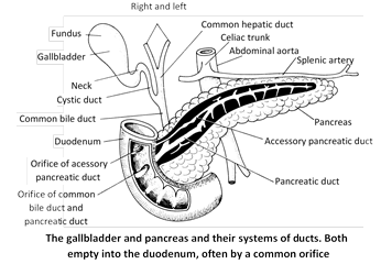
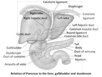
Pancreas : Single, endodermal, flat, leaf-like yellowish, heterocrine (mixed) gland, present between the ascending and descending limb of duodenum and opens into duodenum through pancreatic duct. It’s can devided into following parts-
Exocrine : It is the major part (about 99%) of pancreas. The exocrine tissue of the pancreas consists of rounded lobules (acini) that secrete an alkaline pancreatic juice. The juice is carried by the main pancreatic duct, also called duct of Wirsung, into the duodenum through the hepatopancreatic ampulla (ampulla of vator). An accessory pancreatic duct, also named duct of Santorini, may sometimes lead directly into the duodenum.
Endocrine : Minor part (1% only) also called as islets of Langerhans scattered in the exocrine part. It consist of four various type of cells, as \[\alpha (A)\] cells, \[\beta (B)\] cells, \[\delta (D)\] cells and F or PP cells. \[\alpha -\]cells secretes glucagon hormone, \[\beta -\]cells secretes insulin hormone and \[\delta \] cells secrets somatostatin. The PP or \[F-\]cells secrete pancreatic polypeptid hormone to control somatostatin. The secretion passes directly into blood.
(1) Amount : 1-1.5 l/day
(2) Chemical nature : alkaline
(3) \[pH:\text{ }7.1-8.2\]
(4) Control of secretion : Hormonal and normal mechanism.
Secretin hormones stimulate the production of more alkaline pancreatic juice but low in enzyme content. Pancreozymin or Cholecystokinin stimulates the production of enzyme rich pancreatic juice.
(5) Chemical composition : Water (99%), enzymes and salts.
Functions of pancreas and its enzymes
(1) The islets of Langerhans secrete insulin and glucagon hormones.
(2) The exocrine part of pancreas secretes pancreatic juice.
(3) Elastase : It act upon elastin protein.
(4) Trypsinogen \[\underset{\text{Intestinal}\,\,\text{juice}}{\mathop{\xrightarrow{\text{Enterokinase}\,\,\text{of}}}}\,\] Trypsin.
(5) Trypsinogen \[\underset{\text{(Autocatalysis)}}{\mathop{\xrightarrow{\text{Trypsin}}}}\,\] Trypsin.
(6) Chymotrypsinogen \[\underset{\text{Autocatalysis}}{\mathop{\xrightarrow{\text{Trypsin}}}}\,\] chymotrypsin.
(7) Polypeptides + peptones \[\underset{\text{(Pancreatic}\,\,\text{protease)}}{\mathop{\xrightarrow{\,\,\,\,\,\,\text{Trypsin}\,\,\,\,\,\,}}}\,\] Tripeptides + Dipeptides + Oligopeptides.
(8) Starch \[\underset{\text{(Pancreatic}\,\,\text{amylase)}}{\mathop{\xrightarrow{\text{Amylopsin}}}}\,\] Maltose + Isomaltose + limit dextrin.
(9) Emulsified Lipids \[\overset{\text{Steapsin}}{\mathop{\xrightarrow[\text{(Pancreatic}\,\,\text{lipase)}]{}}}\,\] Fatty acids + Glycerol + Monoglycerides.
(10) Nucleic acid \[\xrightarrow{\text{Nuclease}}\] Nucleotides + Nucleosides.
(11) Nucleic acid \[\xrightarrow{\text{Nucleasidase}}\] Purines + Pyrimidines.
(12) Polypeptides \[\xrightarrow{\text{Chymotrypsin}}\] Oligopeptides.
Liver
(1) Structure : The liver is largest and heaviest gland in the body. Its upper and anterior surfaces are smooth and curved to fit the under surface of the diaphragm; the posterior surface is irregular in outline. It consists of three lobes in frog: right, left and median; five lobes in rabbit: left lateral, left central, spigelian, right central and caudate; four lobes in man: right, left, quadrates and caudate lobe. It is divided into two main lobes : right and left lobes separated by the falciform ligament.
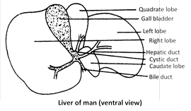
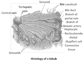
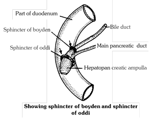
A pear-shaped sac, the gall bladder is attached to the posterior surface of the liver by connective tissue. The right and left hepatic ducts join to form the common hepatic duct. The latter joins the cystic duct, which arises from the gall bladder. The cystic duct and common hepatic duct join to form common bile duct or ductus cholidochus which passes downwards posteriorly to join the main pancreatic duct to form the hepatopancreatic ampulla (ampulla of Vater). The ampulla opens into the duodenum. The opening is guarded by the sphincter of Oddi. The sphincter of Boyden surrounds the opening of the bile duct before it is joined with the pancreatic duct. The basic structural and functional unit of the liver is the hepatic lobule.
Each lobule is composed of plates of polyhedral, glycogen-rich cells, the hepatocytes, arranged radially around a central vein. Between the plates are radial blood sinusoids. At the periphery of the lobules, the branches of portal vein, hepatic artery, bile ducts, and lymphatics course together. A network of tubular spaces between the hepatocytes represents the bile canaliculi. At the periphery of the lobule the bile canaliculi empty into small hering’s canals walled by cuboidal epithelium. These canals lead into bile ducts walled by columnar epithelium. The sinusoids are lined by incomplete endothelium with scattered phagocytic Kupffer cells, that eat bacteria and foreign substances
Gall bladder : The gall bladder is a slate-blue, pear-shaped sac connected with an supported from liver by a small omentum or ligament. Its distal part is called fundus, while the narrow part, continued as cystic duct, is called the neck.

Functions of liver : Liver, the largest gland of vertebrate body, is an essential organ, which performs many functions –
(1) It secretes bile which is a complex watery fluid containing bile salts (Na taurocholate and Na glycocholate), bile pigments (biliverdin and bilirubin), cholesterol, mucin, lecithin and fats etc. It breaks and emulsifies the fat.
(2) In the liver, haemoglobin of the worn out erythrocytes breaks down to bile pigments bilirubin and biliverdin. The bile pigments are also converted in the bowel into stercobilin which colours the faeces.
(3) Excess quantities of carbohydrates (glucose) are converted to glycogen (Glycogenesis) in the presence of insulin in the liver cells, and stored therein.
(4) Glycogen is a reserve food material, which is changed into glucose (Glycogenolysis) and released into the blood at concentrations maintained constant by the liver. In this way, blood–sugar level is maintained under diverse dietary conditions.
(5) Under abnormal conditions, liver can convert proteins and fats into glucose by complex chemical reactions. Formation of this “new sugar” i.e. from non–carbohydrate sources, is called gluconeogenesis.
(6) If the level of blood–glucose rises beyond normal even after glycogenesis and catabolism, the excess glucose is converted into fat and stored in the liver. The process is termed lipogenesis.
(7) Amino acids resulting from protein digestion finally come into the liver from the intestine. They are partly released into the blood for distribution and protein synthesis, partly transaminated into other amino-acids and deaminated.
(8) In the embryo, red blood cells are manufactured by the liver. In the adult, liver stores inorganic salts of iron, copper and vitamin \[{{B}_{12}}\](anti–anaemic factor) and thus helps in the formation of red blood cells and haemoglobin.
(9) Liver functions as a store–house for blood and regulates blood–volume.
(10) Fibrinogen, prothrombin and certain other blood coagulation factors are formed in the liver. Heparin is an intravascular anticoagulant that is stored in the liver.
(11) The plasma proteins serum albumin and serum globulin are synthesized by the liver from the amino acids.
(12) Liver synthesizes vitamin A from the provitamins A (carotenoid pigments). Liver cells also store fat–soluble vitamins A, D, E and K. Besides, it is the principal storage organ for vitamin \[{{B}_{12}}.\]
(13) The liver is the site of detoxification of different toxic substances either produced in the body or taken along with food.
(14) It is the main heat producing organ of the body.
(15) Kupffer cells in the liver sinusoids phagocytose and remove bacteria, worn-out blood elements and foreign particles.
(16) Liver is an important site of lymph formation.
Bile/chole
(1) Amount : \[800-1000\,\,\,\,ml\]daily. On the average about \[700\,\,ml.\]
(2) Source : Secreted by hepatic cells
(3) Storage site : Gall bladder
(4) Colour : Greenish-blue
(5) Chemical nature : Alkaline
(6) \[pH:\text{ }7.68.6\]
Functions of bile
(1) Emulsification of fats.
(2) Helps in absorption of fat-soluble vitamins.
(3) Increases alkalinity to make the medium suitable for enzymatic action.
(4) Elimination of heavy metals such as \[Cu,\text{ }Hg,\text{ }Zn\] etc.
(5) Elimination of excess of bile pigments.
(6) Stercobilin and urobilin (urobilin found in urine) is formed by bilirubin and biliverdin is responsible for colouration of faeces..


You need to login to perform this action.
You will be redirected in
3 sec

