Parts Of Nervous System
Category : 11th Class
Nervous system is divided into three parts -
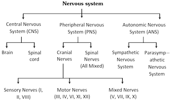
Central nervous system (CNS)
In all the vertebrates including man, CNS is dorsal, hollow and non-ganglionated while in invertebrates when present, it is ventral, solid, double and ganglionated. CNS is formed of two parts :
(1) Brain - Upper and broader part lying in the head.
(2) Spinal cord - Lower, long and narrow part running from beginning of neck to trunk. CNS is covered by 3 meninges and its wall has two type of matter.
Types of matter : CNS of vertebrates is formed of two types of matter –
(i) Grey matter : It is formed of cell-bodies, non-medullated nerve fibres, neuroglea, dendrites of association neurons and motor neurons.
(ii) White matter : It is formed of medullated nerve fibres or myelinated axon of motor and sensory neurons, which appear white due to presence of medullary sheath.
Meninges : The meninges are connective tissue membranes which surround the brain and spinal cord of CNS. In the fishes, there is only one meninx called meninx primitiva (piamater). In amphibians, reptiles and birds, the brain is covered by two meninges or membranes : inner pia-arachnoid and outer duramater. In mammals, CNS is covered by three meninges or membranes or cranial meninges. Brain meninges are continuous with spinal meninges
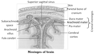
The three layers of cranial meninges in order from superficial to deeper duramater, arachnoid and piamater. Duramater is nonvascular, tough made up of fibrous connective tissue. Arachnoid mater made up of reticular connective tissue with collagen and elastin fiber, while innermost vascular piamater (nutritive) made up of loose aerolar connective tissue. Between dura and arachnoid mater presence of sub dural space (no CSF in mammals here), between Arachnoid and piamater presence of sub-arachnoid space (with CSF in mammals, CSF also found in ventricles and central canal). Between duramater and periosteum presence of epidural space. An extension of duramater between two cerebral hemispheres called falx cerebri. Tentorium, an extension of duramater between cerebrum and cerebellum.
Cerebrospinal fluid : All the ventricles of the brain, central canal of spinal cord are continuous and lined by a columnar, ciliated epithelium, the ependyma. They contain lymph-like extracellular fluid called the cerebrospinal fluid (C.S.F.). This fluid is secreted by the choroid plexuses by filtration of blood. The choroid plexuses consist of loose connective tissue of pia mater covered internally by a simple cuboidal epithelium of secretory (glandular) nature. The cerebrospinal fluid slowly flows toward the fourth ventricle by secretion pressure and passes into the spinal cord. Some fluid escapes into the subarachnoid spaces through three pores a median aperture (of magendie) and a paired lateral aperture (of Luschka) in the roof of the fourth ventricle in the medulla. From the subarachnoid spaces, the cerebrospinal fluid is transferred to the blood of the venous sinuses. Nervous tissue is without lymphatic vessels.
The cerebro-spinal fluid (CSF) provides -
(i) Protection to brain from mechanical shocks, physical injury.
(ii) Optimum physiological fluid environment for neural functions e.g. conduction of nerve impulses, transport of aminoacids, sugars, etc.
(iii) ‘Relief’ mechanism for the increase in intracranial pressure that occurs with each arterial pulse of blood to brain.
(iv) ‘Sink’ like facility for metabolites of brain.
(v) The blood CSF barrier for selective transport process between blood and CSF.
(vi) Nourishment to CNS.
Major site of CSF formation is choroid plexus, and mid ventricular wall and sub-arachnoid wall also contribute. CSF is cell free, slightly alkaline, and is isotonic to plasma. Rate of formation of C.S.F is 20 ml/h (480 ml/day) 20 ml/hour approx, 1/2 litre per day. Total amount present in and around CNS is 80-150 ml it means there is atleast 3 times renewal of C.S.F. every day. CSF contains glucose, proteins, lactic acid, urea, and some WBC.
Blood brain barrier facilitate maintenance of stable internal environment. Its acts as physiological and pathological barrier.
There are three choroid plexus in humans –
(i) Lateral choroid plexus : It is in the roof of I and II ventricle.
(ii) Anterior choroid plexus : It is in the roof of III ventricle (diacoel).
(iii) Posterior choroid plexus or pelochoroida : It is in the roof of IV ventricle.
Oxygen and glucose requirements : Brain controls the functions of our body organs and also provides the qualities of mind -learning, reasoning, and memory. For these activities, brain needs a large and constant energy supply. At any given time, the activities of the brain account for 20% of the body’s consumption of oxygen and 15% of its consumption of blood glucose. Brain deprived of oxygen for just 5 minutes is permanently damaged. Mental confusion results if brain is deprived of glucose.
Structure of human brain (Encephalon)
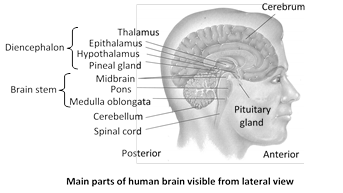
It is soft, whitish, large sized and slightly flattened structure present inside cranial cavity of cranium of the skull. In man, it is about 1200-1400 gm in weight and has about 10,000 million neurons. Brain is made up of 3 parts :
(1) Fore brain or Prosencephalon : It forms anterior two-third of brain and is formed of three parts.
Olfactory lobes : These are one pair, small sized, club-shaped, solid, completely covered by cerebral hemisphere dorsally. Each is differentiated into two parts -
(i) Olfactory bulb : Anterior, swollen part, and
(ii) Olfactory tract : Posterior and narrow part which ends in olfactory area of temporal lobe of cerebral hemisphere.
Function : These control the smell.
(a) It is normal in frog, rabbit and man.
(b) It is well developed in dog. So power of smell is more in dog.
(c) These are also well developed in dog fish and name dog fish is on the basis of well developed olfactory lobes.
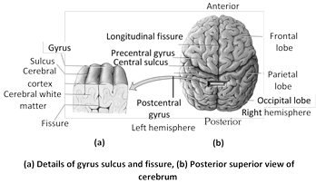
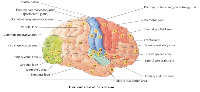
Cerebrum : Cerebrum is divided into 5 lobes (a) frontal (b) parietal, (c) occipital, (d) temporal and (e) Insula. A lobe called insula is hidden as it lies deep in the sylvian fissure. The cerebral hemisphere are separated from olfactory lobes by rhinal fissure. The median fissure divides the cerebrum into a right and a left cerebral hemisphere.
A few sulci are well developed and form three deep and wide fissures which divide each cerebral hemisphere into four lobes : anterior frontal lobe, middle parietal lobe, posterior occipital lobe and lateral temporal lobe e.g. Fissure sulcus lying between the frontal and parietal lobes is central fissure or sulcus, that lying between the parietal and occipital lobes is parieto-occipital fissure and that demarcating frontal and parietal lobes from the temporal lobe is lateral or Sylvian fissure. Each cerebral hemisphere is with a fluid-filled cavity called lateral ventricle or paracoel.
Two cerebral hemispheres are interconnected by thick band of transverse nerve fibres of white matter called corpus callosum. The peripheral portion of each cerebral hemisphere is formed of grey matter and is called cerebral cortex, while deeper part is formed of white matter and is called cerebral medulla. Cerebral cortex is the highest centre for many sensations and activities and is with a number of sensory areas. Cerebral cortex 2-4 mm thick.
Two cerebral hemispheres are interconnected by thick band of transverse nerve fibres of white matter called corpus callosum. The peripheral portion of each cerebral hemisphere is formed of grey matter and is called cerebral cortex, while deeper part is formed of white matter and is called cerebral medulla. Cerebral cortex is the highest centre for many sensations and activities and is with a number of sensory areas. Cerebral cortex 2-4 mm thick.
Histology of cerebrum : The whole brain possess grey matter outside and white matter inside around ventricle.
(i) Grey matter : In cerebrum grey matter is very much developed, it is on an average 2-4 mm. thick but at poles its thickness is 1.3 mm. It is thickest at pre central gyrus (4.5 mm thick). Grey matter of cerebrum is called cortex or pallium. Phyllogenetically or evolutionarily cortex is divided into 3 parts -
(a) Allocortex or paleocortex : It is the cortex of olfactory area of frontal lobe and olfactory bulbs. In lower vertebrates (cartilagenous fish) olfactory lobes occupy most of the part of cerebrum. So in these animals sense of olfection is very-very much developed. Sense of olfaction is oldest sense.
(b) Mesocortex : It is relatively not much older in development.
(c) Neocortex or neopallium or isocortex or neencephalon : It is most recent cortex and is developed maximum only in human. It is in prefrontal cortex or prefrontal region (organ of mind), precentral and precentral gyrus etc. The neocortex is having 6 layer of neurons while remaining cortex possess only 5 layers.
The cerebral cortex is having area of about 2200 cm2 while the cranial cavity is only 1450 cm3, so to accomodate cerebrum there appears foldings in the cortex. The ridges are called gyrus (or gyri) or convolution while the depression are called sulcus (sulci in plural).
(ii) White matter : It is inner part of brain. White matter is aggregation of myelinated and unmyelinated axons of many neurons. Its fibres are divide into 3 categories :
(a) Commissural fibers : These neurons connect gyri of 2 hemispheres, such as corpus callosum. habenular commissure, anterior commissure, posterior commissure.
(b) Associate fibres : They connect gyri of same hemisphere.
(c) Projection neuron : They are infact ascending and descending nerve tract, they connect one part of brain to another part of brain or to spinal cord. (In spinal cord they were called as columo).
Associated structures of cerebrum : Cerebrum has following specific structure.
(i) Sub cortex : Nuclei on white matter. It is cluster of grey neurons in depth of white matter, they are formed in whole brain and are named differently.
(ii) Basal ganglia or central nucleus : These are several groups of nuclei in each cerebral hemisphere.
Corpus striatum : Corpus striatum is the largest nucleus, consist of caudate nucleus and lenticular nucleus. The lenticular nucleus is sub-divided in putamen (outer shell) and globus pallidus (ball). Other structure, functionally linked to and some times considered part of basal ganglia are :
(a) Claustrum : It is the name given to grey matter present between insula and putamen.
(b) Epistriatum or Amygdaloid body : It is structure present at the end of caudate nucleus.
(c) Red nucleus and substantia nigra of mid brain.
(d) Sub thalamic nuclei of diencephalon.
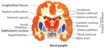
Function of basal ganglia
(i) Caudate and putamen control large automatic movements of skeletal muscle like swinging of arm while walking.
(ii) Globus pailidus control muscle tone for specific body movements.
(iii) Corpus callosum : It is the band of white neurons present between both cerebral hemisphere and connect them on medial surface. It is present only mammal. It has anterior part genu, middle part trunchus and last part splenium.
Below corpus callosum there are two fused band of white neurons called fornix. There anterior part is called column and posterior part is called crura. Between column and genu a membrane is called septum lucidum or septum pellicidum. Septum lucidum encloses a space called V5 or Pseudocoel, because it is not possessing C.S.F. i.e. why it is called pseudocoel.
(iv) Limbic system : Limbic system present on inner border of cerebrum and floor of diencephalon, It is also called emotional brain or animal brain. Limbic system controlling emotion, animal behaviour like chewing, licking, sniffing, docility, tameness, affection (animals) rage, pain, pleasure, anger, sexual feelings, feer, sorrow grooming. It has following structure -
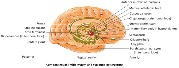
(a) Cingulate gyrus : It is a region of pre central gyrus.
(b) Hippocampal gyrus : It is a region of temporal lobe near colossomarginal sulcus. These two structure are combinely called limbic lobe.
(c) Amygdaloid body : It is the end of caudate nucleus.
(d) Olfactory bulb : They are on the inferior anterior surface of brain. Olfactory nerve ends in these bulb.
(e) Mammillary body : They are found in hypothalamus. Olfactory bulb and mammillary body both are centre of olfaction.
(f) Dentate gyrus : Is in between hippocampus and parahippocampale.
(g) Anterior nucleus of thalamus is located in floor of lateral ventricle.
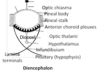
Diencephalon : Diencephaton cavity is called, III ventricle or diocoel the thin roof of this cavity is known as the epithalamus, the thick right and left sides as the thalami, and floor as the hypothalamus.
(i) Epithalamus : It forms roof of third ventricle. The epithalamus is not formed of nervous tissue. It consists of piamater only. Hence, it is of relatively little significance as a nerve centre. Its anterior part is vascular and folded. It is called anterior choroid plexus. Behind this plexus, the epithalamus gives out a short stalk, the pineal stalk which bears a small, rounded body, the pineal body, at its tip, and paired right and left habenular nuclei (olfaction or smell).
(ii) Thalamus : A pair of mass of grey matter forms the superior part of lateral walls of the third ventricle. It measures 3 cm in length and comprises 80% of diencephalon. The thalamus is principal relay station for sensory impulses that reach the cerebral cortex from spinal cord, brain stem, cerebellum. It also allows crude appreciation of some sensations such as pain, temperature, and pressure. Certain nuclei in the thalamus relay all sensory input to cerebral cortex. These include the -
(a) Medial geniculate nucleus for hearing.
(b) Lateral geniculate nucleus for vision.
(c) Ventral posterior nucleus for taste, touch, pressure, vibration, heat, cold, and pain.
Other nuclei are centers for synapse in somatic motor system their include.
(a) Ventral lateral nucleus and ventral anterior nucleus (voluntary motor actions).
(b) Anterior nucleus concerns with emotions and memory.
(iii) Hypothalamus : The hypothalamus is visible in the ventral view of the brain and forms the floor of diencephalon. Hypothalamus also gives a nervous process called infundibulum (forms pars nervosa) which meets a rounded non-nervous pharyngeal outgrowth called hypophysis. Both collectively form master gland called pituitary body. A stalked outgrowth of infundibulum combines with a pouch-like epithelial outgrowth (Rathke’s pouch) of the roof of embryonic mouth (= stomodaeum), forming a pituitary gland or hypophysis. Which secretes a number of hormones. In front of hypothalamus, there is cross of two optic nerves called optic chiasma. Behind the hypothalamus, there is one pair of small, rounded, nipple-like bodies called mammilary bodies or corpora mammillares. The hypothalamus consists of many masses of grey matter, called hypothalamic nuclei, scattered in the white matter.
In man and some other mammals, most fibres of optic nerves cross, but some fibres do not cross and innervate the eyes of their own respective sides. This arrangement enables man and these mammals to have a binocular vision. Rabbits simply have a monocular vision.
Pineal gland is a pine cone-shaped gland. It is located in the center of brain with which it loses all nerves connection after birth. It is innervated by sympathetic nerves. It has a photosensory role in amphibian and primitive reptiles and is called ‘Third eye’. Pinealocytes secretes melatonin. Mammalian pineal does not act as photoreceptor but it produces the hormone called melatonin which is anti FSH, and anti LH. It inhibits reproductive function. Melatonin secretions decrease after puberty.
Functions of fore brain
(i) Olfactory lobe : It is centre of smell.
(ii) Cerebrum : Cerebral cortex is made up of grey matter and differentiated into –
(a) Sensory and associated area confirm, recognise and evaluate for shape, colour, sound, taste and smell for sensory cells in relation with object.
(b) Broca’s area : Known as sensory speech area or motor speech area. Translate thought into speech. Located into frontal lobe towards left side. It is associated with language area and also interpriate translation of written words into speech. Damage or injury in Broca’s area (sensory or motor speech area) may result
Aphasia(Inability to speak), Word deafness, Word blindness.
Important areas in the human brain
|
Area |
Location |
Function |
|
Premotor area |
Frontal lobe |
The highest centre for involuntary movements of muscles and ANS. |
|
Motor area |
Frontal lobe |
Controls voluntary movements of specific the muscle |
|
Broca?s area |
Frontal lobe |
Motor speech area (Translation of thought and written words into speech) |
|
Somesthetic area |
Parietal lobe |
Perception of general sensation like pain, touch and temperature |
|
Auditory area |
Temporal lobe |
Hearing (Interprets characteristics of sound such as pitch and rhythm. |
|
Olfactory area |
Temporal lobe |
Sense of smell |
|
Wernicke?s area |
Temporal lobe |
Understanding speech written and spoken |
|
Gustatory area |
Parietal lobe |
Sense of taste |
|
Visual area |
Occipital lobe |
Sensation of light |
(c) Cerebrum is a centre for -Intelligence, Emotion, Will power, Memory, Consciousness, Imagination, Experience, Knowledge, Reasoning, Voluntary controls, Weeping and laughing, Micturition, Defecation.
(iii) Diencephalon is a centre for :
(a) Carbohydrate metabolism
(b) Fat metabolism
(c) It relays impulses from posterior region of brain and also to posterior region of brain.
(d) Its secretes neurohormone
(e) From part of pituitary gland
(f) Secrete cerebrospinal fluid
(iv) Hypothalamus is a centre for -Hunger, Thirst, Sweating, Sleep, Fatigue, Temperature, Anger, Pleasure, love and hate, Satisfaction.
(2) Mid brain or mesencephalon : Extends from pons to diencephalon, contain both white and gray matter. Posterior portion of mid brain called tectum. It is also completely covered by cerebral hemisphere. It is formed of two parts -
(i) Optic lobes : These are one pair, large sized lobes present on dorsal side. Each is divided transversely into upper and larger superior coliculus and lower and smaller inferior coliculus. So there are four optic lobes, so called optic quadrigemina (only in mammals). In frog these are known as bigemina. Valve of vieussens. It joins the optic lobe with cerebellum.
(a) Superior optic lobe or superior colliculus : They are concerned with reflex action of eye, head and neck in response to visual stimulus.
(b) Inferior colliculus : They are concerned with movement of head and trunk in response to hearing stimulus.
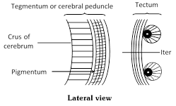
(ii) Cerebral peduncle (crura cerebri) : They are the pair of thick bands of longitudinal nerve fiber present on the floor or ventral side of mid brain. The dorsal part of cerebral peduncle (white matter) is called Tagmentum while most ventral part (gray matter) is called crura cerebrae or crus of cerebrum. Dorsal thick wall of mid brain is known as optic tectum. Iter is between tegmentum and tectum. Cerebral peduncle are infect possessing ascending and descending tracts, connecting upper and lower region of brain.
In white matter of cerebral peduncle these are following sub cortical structure
(a) Red nucleus or rustrum nucleus : They are red because rich blood supply and iron containing pigment or haemoglobin. Function with basal ganglia and cerebellum to coordinate muscular movement.
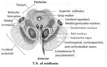
(b) Substantia nigra : It is black because of much deposition of melanin.
(c) Occulomotor nucleus : It is origin point of 3rd cranial nerve (occulomotor) from this region 4th (Trochlear) nerve also originates.
Functions of Mid brain
(i) Pair of anterior optic lobes (which are also known as superior colliculi) is related with vision.
(ii) Pair of posterior optic lobe (known as inferior colliculi) related with auditory.
(iii) These act as coordination centres between hind and fore brain.
(3) Hind brain : Hind brain consists of (i) cerebellum and (ii) medulla oblongata (iii) Pons varolii.
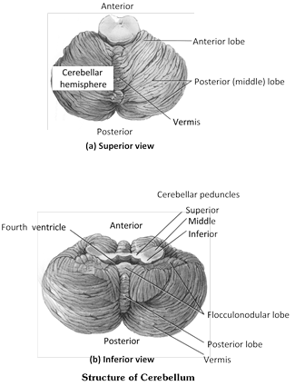
(i) Cerebellum (Sandwitched brain) : Cerebellum is second largest portion of brain lies posterior to medulla and pons and inferior to posterior portion of cerebrum. Cerebellum separated by cerebrum by a transverse fissure and by an extension of cranial dura mater called tentorium cerebelli. Cerebellum is butterfly shape consist of :
(a) The central constricted area is vermis
(b) A lateral wings or lobes called cerebellar hemispheres with anterior and posterior lobe (govern skeletal muscle movement).
(c) The flocculo nodular lobe (sense of equilibrium).
Between cerebellar hemisphere is extension of cranial dura mater called falx cerebelli. The superficial layer of cerebellum, called cerebellar cortex, consist of gray matter in series of parallel ridges called folia. Deep to gray matter are white matter tree called ‘Arbor vitae’ or tree of life. Cerebellum attached to brain stem by three paired cerebellar peduncles -
(a) Inferior cerebellar peduncle between cerebellum and medulla with sensory/motor fiber.
(b) Middle cerebellar peduncle between cerebellum and pons with sensory axon.
(c) Superior cerebellar peduncle between cerebellum and mid brain, mainly with major fiber.
Cerebellum receives sensory impulses from proprioceptors in muscle, joint, and tendons, coordinate skeletal muscle contractions and also regulate posture and balance.
(ii) Medulla oblongata
Medulla oblongata is the hindest and posterior most part of brain. Cavity is known as IVth ventricle (metacoel). Which is continuous with central canal of spinal cord. It has a pair of lateral Foramina of Luschka and a median foramen magendie. Cerebrospinal fluid come in contact by these apertures from internal cavity of the brain to outer fluid of meninges. A arrangement on its ventral surface there are buldgings of ascending and descending tract which are called pyramids. On the ventral surface these pyramids cross each other which is called decussation of pyramids. On the dorsal side of medulla there are two nuclei which are called nucleus gracilis (long) and nucleus cuneatus. On floor of there is groove called calamus scroptosious.
In the medulla oblongata, most of the sensory and motor fibres cross from one side to the other. Thus, the left cerebral hemisphere controls the right side of the body and vice versa. The reason for this is not known. The lower end of medulla passes into the spinal cord. There is no demarcation between the two. However, the medulla is considered to start at the level of the foramen magnum of the cranium. Medulla contain nuclei of origin of 5 pairs of cranial nerves, VIII, IX, X, XI and XII. VIII -vestibulocochlear nerve concerning with hearing and equilibrium. (There are also nuclei for vestibular branch of VIII in pons).
(iii) Pons Varolii : An oval mass, of white mater called the pons varolii, lies above the medulla oblongata. It consists mainly of nerve fibres which interconnect as bridge connecting spinal cord with brain and parts of brain with each other. Pons also with pneumotaxic area and apneustic area. Together with medullary rhythmicity area, they help control respiration.
Cerebellum -
(i) Poorly developed in frog but well developed in mammal.
(ii) It is centre for co-ordination of muscular movement.
(iii) It is primary centre for balancing, equilibrium, orientation.
Medulla oblongata contain centre for -
(i) Heart beats
(ii) Respiration
(iii) Digestion
(iv) Blood pressure
(v) Gut peristalsis
(vi) Swallowing of food
(vii) Secretion of gland
(viii) Involuntory function -e.g. vomiting, coughing vasoconstrictor, vasodilater, sneezing, hiccouping.
(ix) It control urination, defecation.
(x) The cardiovascular center -regulate rate, force of heart beats.
(xi) Medullary rhythmicity area -adjust basic rhythm of respiration.
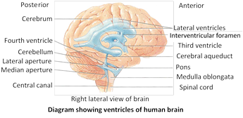
Cavities or ventricles of the brain
The ventricles consist of four hollow fluid filled space inside the brain and same duct for connection between these ventricle.
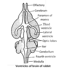
(1) Olfactory lobe -Rhinocoel
(2) Cerebrum -I and II ventricle or lateral ventricle or paracoel.
(3) Foramen of monero : I and II ventricle communicating with IIIrd ventricle by foramen of monero. They are two in human and single in rabbit and frog.
(4) Diencephalon : Third ventricle or Diocoel.
(5) Iter or cerebral aquiduct or aquiduct of sylvius : It is very narrow cavity between III and IV ventricle.
(6) Optic lobe : Optocoel
(7) Cerebellum : Solid.
(8) Medulla oblongata : 4th ventricle or metacoel.
Cavities of brain and spinal cord are modified neurocoel. They are lined by low columnar ciliated epithelium called ependyma.
Subdivisions, parts and associated structures of a vertebrate brain
|
Divisions |
Subdivisions |
Parts |
Cavity |
Associated strcutures |
|
|
(1) Telencephalon |
Rhinencephalon |
I Ventricle (Rhinocoel) |
Olfactory bulbs, Olfactory tracts, Olfactory lobes, Palaeocortex on pallium |
|
|
Cerebral hemispheres |
II or Lateral Ventricles
|
Corpora striata or basal ganglia, Corpus callosum, Neocortex on pallium, Paraphysis |
|
|
(I) Prosencephalon (Forebrain) |
(2) Diencephalon |
Epithalamus (roof) |
|
Habenulae, Pineal apparatus, Parapineal or parietal |
|
|
Thalamus (sides) superior |
|
|
|
|
Hypothalamus (floor), Inferior side |
|
Hypothalamic nuclei, Optic chiasma, Median eminence, Infundibular stalk, Pituitary, Saccus vasculosus, Mamillary bodies, Anterior choroid plexus |
||
|
(II) Mesencephalon (Midbrain) |
- |
Crura cerebri (floor) |
Iter or cerebral aqueduct |
Corpora quadrigemina (superior colliculi, inferior colliculi), Tectum, substantia nigra and red nuclei. |
|
(III) Rhombencephalon (Hind brain) |
(1) Metencephalon |
Cerebellum |
|
Trapezoid body, Pons |
|
(2) Myelencephalon |
Medulla oblongata |
IV Ventricle (Metacoel) |
Restiform bodies, Pyramids |
Salient or mammalian features of human brain : The salient or mammalian features in the human brain are -
(1) Relatively small, solid olfactory lobes.
(2) Very large cerebral hemispheres divided into lobes and with highly folded surface, with cerebral cortex of gray matter.
(3) Corpus callosum interconnecting the cerebral hemispheres only found in eutheria.
(4) Very small pineal body.
(5) A pair of mammillary bodies joined to hypothalamus.
(6) Relatively small, solid optic lobes divided into 4 corpora quadrigemina.
(7) Large, solid cerebellum, with highly folded surface and divided into lobes.
(8) Pons varolii present anterior to the cerebellum.
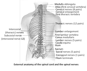
Spinal cord
Present in spinal canal or vertebral canal of vertebral column. It is extended from foramen magnum to II lumber vertebra. In new born infants, extend to 3 or 4 lumber vertebra. Spinal cord is swollen in cervical and lumber region which are called cervical and lumber enlargement. The length of spinal cord ranges from 42 to 45 cm. Its diameter is about 2cm.
(1) Conus medullaris : It is last tapering ends of spinal cord, its ciliated central canal is called Vth ventricle.
(2) Cauda equina : A horse tail-like collection of roots of spinal nerves at inferior end of the spinal cord. Some spinal nerves arise from inferior part of cord do not leave vertebral column at some level as they exit from spinal cord. The roots of these nerves angle inferiorly in vertebral canal from end of spinal cord like wisps of hair.
(3) Filum terminales : It is extension of piamater below conus medullaries up to coccyx. In frog spinal cord also extends upto end of vertebral column.
(4) Cisterna terminalis : It is last dilation of subarachnoid space below 1st lumbar vertebra. It is a proper site for lumber puncture or spinal tap, which is done to drain C.S.F out (5 to 10 ml). This C.S.F is used in diagnosing many diseases of CNS like meningitis, cyphalis, inter cranial pressure, menningococcal inferaction etc.
(5) Meninges : Like brain, spinal cord is also enclosed with in three membranes. In this case duramater does not remain attached with the vertebra, instead there is a space between duramater and vertebra called epidural space. The epidural space is filled with a fluid. The distribution of duramater and piamater in spinal cord is the same as that of brain.
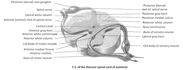
The cross section of spinal cord reveals the following structures -
(1) Central canal : In the centre of spinal cord, there is a canal called central canal. It is filled with cerebrospinal fluid. central canal is continuous with 4th ventricle of medulla oblongata.
(2) Dorsal fissure : In the mid dorsal line, there is a groove extending throughout its length.
(3) Ventral fissure : It is also a groove situated in the mid ventral line throughout the length of spinal cord.
(4) Dorsal septum : It is a partition extending from dorsal fissure to central canal.
(5) Grey matter : It lies around the central canal in the form of a butterfly and sub-divided into 3 horns, surrounded by white mater.
(6) Dorsal horns : It is like horn of grey matter on the dorsal side.
(7) Ventral horns : On the ventral side of the grey matter are horn like structures the ventral horns.
(8) Lateral horns : These are horns on the lateral side of grey matter.
(9) White matter : White matter is present around grey matter. Dorsal and ventral horn, divide white mater into 3 broad area on each side -
(i) Anterior (ventral) white columns
(ii) Posterior (dorsal ) white columns
(iii) Lateral white columns.
Reflex action : Reflexes are fast predictable, automatic responses to change the environment. First of all Marshal Hall (1833) studied the reflex action. Best and Taylor defined reflex action “simplest form of irritability associated with the nervous system is reflex actions or a reflex reaction is an immediate involuntary response to a stimulus.” The reflex actions are involuntary actions because these are not under the conscious control of the brain. Central nervous system is responsible for the control of reflex action.
Reflex arc is formed by the neurons forming the pathway taken by the nerve impulses in reflex action. The simplest reflexes are found in animals involving a single neuron and the following pathway -
\[\text{Stimulus }\ \to \ \,\text{Receptor}\ \xrightarrow{\text{Neuron}}\,\ \text{Effector}\ \,\to \,\ \text{Response}\]
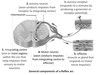
(1) Component of reflex action : The whole of the reflex are includes five parts -
(i) Receptor organs : Receptors are windows of the body or guards of the body. These are situated on all, important organs, for example -eyes, nose, ear, tongue, integument etc. These perceive the stimuli from out side the body.
(ii) Sensory neurons : These are also termed afferent neurons. These carry the stimuli from receptors to spinal cord. These neurons are situated in the ganglion on the dorsal side of spinal cord gray matter or brain stem.
(iii) Association neurons or Integrating center : These are also called intermediate neurons or interstitial neurons. These are found in spinal cord. They transfer the impulses from sensory neurons to motor neurons. Association neurons forms monosynaptic and polysynaptic reflex arc.
(iv) Motor neurons : These are situated in the ventral horn of spinal cord. These carry the impulses to effector organs.
(v) Effector organs : These are the organs, which react and behave in response to various stimuli, for example -muscles and glands.
(2) Mechanism of reflex action : The time taken by a reflex action is too short, for example -in frog it is 0.3 meter per second and in man 5-120 meter per second. Whenever, a part of the body is stimulated by any stimulus, for example -pin pricking, then the stimulus is converted into impulse. This impulse is perceived by the dendrites of sensory neurons. From here, the stimulus reaches the spinal cord through axonic fibres. In the spinal cord, this stimulus passes through synaptic junctions and reaches the intermediate neurons, from where this stimulus reaches the effector organs through motor nerve fibres. As soon as the stimulus reaches the effector organs, it is stimulated and that part of the body is immediately withdrawn. The whole reflex action takes place so rapidly and quickly that we know it when it is completed.
(3) Type of reflexes : The reflexes are of following types –
(i) Monosynaptic reflex : This is the simplest reflex found in vertebrates. The simplest reflex found in vertebrates. The sensory neuron synapses directly on to the motor neuron cell body. In this case the reflex action takes place without the involvement of brain.
(ii) Polysynaptic spinal reflex : This has at least two synapses situated within the spinal cord. It involves a third type of neuron also -the internuncial or inter-mediate relay neuron. The synapses take place between the sensory neuron and intermediate neuron, and between intermediate neuron and the motor neuron. These two reflex arcs allow the body to make automatic, involuntary, homeostatic adjustments, to changes in the external environment, such as the iris pupil reflex and balance during locomotion, and also in the internal environment such as breathing rate and blood pressure.
(iii) Polysynaptic spinal/brain reflexes : In this case the sensory neuron synapses in the spinal cord with a second sensory neuron, which passes to the brain. The latter sensory neurons are part of the ascending nerve fibre tract and have their origin in preintermediate neuron synapse. The brain is capable of identifying this sensory information and stores it for further use. The motor activity may be initiated by the brain anytime and the impulses are transmitted down the motor neurons in descending nerve fibre tract, to synapse directly with spinal motor neurons in the postintermediate synaptic region.
(iv) Simple reflex : Simple reflex is also known as unconditioned reflex. It is inborn, unlearned, reflex to a stimulus. Simple reflex is mostly protective in function. Example of simple reflex are
(a) Knee jerk -Tendon of patella tapped, also called patellar reflex.
(b) Corneal reflex (blinking reflex) -closing of eyelids.
(c) Rapid withdrawal of hand while burned or pricked.
(d) Quick recovery of balance while falling.
(e) Scratch reflex of frog -in pitched frog with acetic acid.
(f) Coughing, sneezing and yawning.
(v) Acquired reflex : Acquired reflex is also known as conditioned reflex. It is not inborn, but acquired and dependent on past experience, training and learning. Demonstration of conditioned reflex was first made by Russian physiologist Ivan Petrovitch Pavlov (1846-1936) in hungry dog. Pavlov rang the bell while feeding dog, thus associated the unconditioned response with additional stimulus. Examples of conditioned reflex are learning of dancing, cycling, swimming, singing,, driving, etc. These actions are under cerebral control during learning.
Peripheral nervous system
It is formed of a number of long, thin, whitish threads called nerves extending between central nervous system and body tissues. Each nerve is formed of bundles of nerve fibres, fasciculi, held together by connective tissue and surrounded by a white fibrous connective tissue sheath called epineurium.
The nerve fibres are classified into two categories on the basis of presence or absence of myelin (white fatty) sheath.
(1) Medullated or Myelinated nerve fibres.
(2) Non-medullated nerve fibres.
On the basis of function, the nerves are of three types
(1) Sensory nerve
(i) It contains only sensory nerve fibres.
(ii) It conducts nerve impulses from sense organs to CNS to produce sensation. e.g. Optic nerve, auditory nerve.
(2) Motor nerve
(i) It contains only motor nerve fibres.
(ii) It conducts nerve impulses from CNS to some muscles or glands to control their activities. e.g. Occulomotor nerve, hypoglossal nerve.
(3) Mixed nerve
(i) It contains both sensory and motor nerve fibres.
(ii) It conducts both sensory and motor impulses. e.g. All spinal nerves, trigeminal nerve.
On the basis of their origin, nerves are of two types
(1) Cranial or cerebral nerves which either arise from or end into brain.
(2) Spinal nerves which arise from spinal cord.
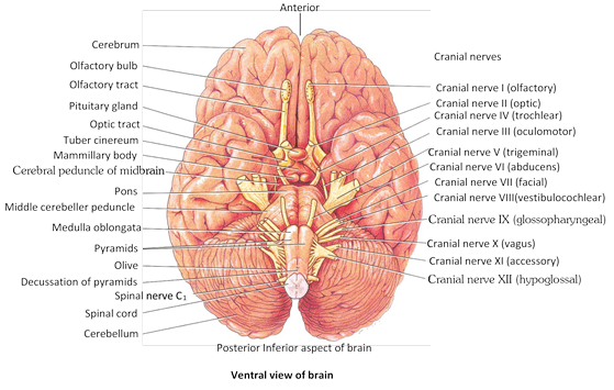
Cranial nerves
(1) 10 pairs of cranial nerves are present in an anamniote (fishes and amphibians).
(2) Number of cranial nerves found in frog is ten pairs (20).
(3) 12 pairs of cranial nervers are present in an amniote (reptiles, birds and mammals).
(4) Number of cranial nerves found in rabbit and man is 12 pairs (24).
(5) The first 10 pairs are common for frog and rabbit. The additional pairs found in rabbit are spinal accessory and hypoglossal.
(6) The smallest cranial nerve is trochlear in human beings, but all animals smallest cranial nerve is abducens.
(7) The largest cranial nerve is trigeminal in human beings but vagus is largest cranial nerve in all animals.
(8) Vagus supplies the regions other than head.
(9) The sensory cranial nerves are
I Olfactory – Smell
II Optic – Vision
VIII Auditory – Hearing and equilibrium
(10) The motor cranial nerves are : III, IV, VI, XI and XII.
(11) Extraocular muscle nerves are : III, IV and VI.
(12) The mixed cranial nerves are : V, VII, IX and X (4 pairs).
(13) Number of cranial nerves in snake (Amniota) 10 pairs.
Cranial nerves of mammal at a glance
|
|
Name |
Nature |
Origin |
Distribution |
Function |
|
(1) |
Olfactory Nerves |
Sensory |
Olfactory lobe |
Sensory epithelium of olfactory sacs |
Receive stimuli from the sensory epithelium of olfactory sac and carry them to olfactory lobes |
|
(2) |
Optic nerves |
Sensory |
In retina of eye |
Lateral geniculate nuclei of thalamus |
Stimulus of light is carried to optic occipital lobe of cerebral cortex. |
|
(3) |
Occulomotor nerves |
Motor |
Crura cerebri (mid brain) |
Eye ball muscles like superior rectus, medial rectus, inferior rectus and inferior oblique. except superior oblique muscle and external rectus |
Movement of eye lids and eye ball. |
|
(4) |
Trochlear nerves |
Motor |
From in between the optic lobes and cerebellum |
Superior oblique muscle of eye ball |
Movement of eye ball |
|
(5) |
Trigeminal nerves |
Mixed |
From the gassarion galglia situated on the lateral side of pons |
- |
- |
|
|
(i) Ophthalmic nerve |
Sensory |
,, |
Skin of lips, upper eye lid, lacrimal, gland |
|
|
|
(ii) Maxillary |
Sensory |
,, |
Upper lip, skin of nose, lower eye lid. Upper teeth. |
Carry the stimuli from these organs to brain |
|
|
(iii) Mandibular nerve |
Mixed |
,, |
Lower lip and skin of jaw |
Carry the stimuli from these organs to brain |
|
(6) |
Abducens nerves |
Motor |
Pons |
Eye muscles external rectus |
Movement of eye ball |
|
(7) |
Facial nerves |
Mixed |
Pons |
- |
- |
|
|
(i) Palatinus |
Sensory |
- |
In the roof of mouth cavity |
Carry the impulses from roof of mouth cavity |
|
|
(ii) Hyomandibular |
Motor |
- |
Muscles of low jaw, muscles of neck and pinna (external ear) |
Carry the impulses from brain muslces of lower jaws, neck and pinna. |
|
|
(iii) Chordotympani |
Mixed |
- |
In salivary glands and taste buds |
Receives the stimuli from the taste buds and carry the stimulus to salivary gland. |
|
(8) |
Auditory nerves |
Sensory |
Medulla, pons |
- |
- |
|
|
(i) Vestibular nerve |
,, |
- |
Semicircular canals, saccule, utricle. |
Receives impulses from the internal ear and carry to brain for equilibrium |
|
|
(ii) Cochlear nerve |
,, |
- |
Cochlea |
Impulses associate with hearing. |
|
(9) |
Glossopharyngeal nerve |
Mixed |
In medulla |
Taste buds present in tongue and muslces of oesphagus |
Secretion of saliva, taste muscle sense (proprioception) |
|
(10) |
Vagus nerve |
Mixed |
Arising from medulla, 9th and 10th cranial nerves unites to form vagus nerve but become separate and divide into branches |
- |
- |
|
|
(i) Superior laryngeal nerve |
Motor |
- |
Glottis, trachea, lung muscle |
(1) Smooth muscles contraction and relaxation. (2) Secretion of digestive juice. (3) Muscle sense (proprioception) (4) Sensation of visceral organs. |
|
|
(ii) Recurrent laryngeal nerve |
Motor |
- |
Glottis, trachea, lungmuscle. |
|
|
|
(iii) Cardiac nerve |
Motor |
- |
Heart Muscles |
From brain to heart muscles |
|
|
(iv) Pneumogastric |
Motor |
- |
In the abdominal cavity, in stomach and lungs. |
Carry impulse from these organs to brain and from brain to muscles of these organs. |
|
|
(v) Depresser nerve |
Motor |
- |
Diaphragm |
Carry the impulse to diaphragm |
|
(11) |
Spinal accessory |
Motor |
Medulla |
Muscles of neck and shoulders, voluntary muscles of pharynx, larynx, and soft palate. |
Swallowing movements, movement of head. |
|
(12) |
Hypoglossal nerve |
Motor |
Medulla |
Muscles of tongue and neck |
Movement of tongue during speech, and swallowing, proprioception (Muscle sense). |
Spinal nerves : Spinal nerves arise from gray matter of spinal cord. There are 31 pairs of spinal nerves in man (37 pairs in rabbit). All spinal nerves are mixed. The spinal nerves in man are divided into 5 groups.
(1) Cervical (C) \[\to \] 8 pairs - in Neck region
(2) Thoracic (T) \[\to \]. 12 pairs - in thoracic region
(3) Lumbar (L) \[\to \] 05 pairs - upper part of abdomen
(4) Sacral (S) \[\to \] 05 pairs - lower part of abdomen
(5) Coccygeal (CO) \[\to \] 01 pairs - represent the tail nerves
Total = 31 pairs
Number of spinal nerves in frog is 10 pairs. In some frog like Rana tigrina, 10th pair may reduced or absent. The first pair of spinal nerves in frog is hypoglossal. The last pair of cranial nerves of mammals has the same name. Brachial plexus is formed by 2nd and 3rd spinal nerves in frog. Sciatic plexus is formed by 7, 8 and 9 spinal nerves in frog. Glands of Swammerdam are calcareous glands found at the places of emerging of spinal nerves in frog.
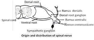
Spinal nerve formula can be written as \[-{{C}_{8}},{{T}_{12}},{{L}_{5}},{{S}_{5}},C{{O}_{1}},\] Spinal nerves exit via intervertebral foramen. Each spinal nerve arises from spinal cord by 2 roots
(1) Dorsal (= Afferent = Sensory = Posterior) root is a continuation of dorsal horn and is formed of gray matter. It presents a ganglionic swelling in middle, called dorsal root ganglion. These transmit sensory nerve impulses from the sense organs to spinal cord (touch, pain, temperature). They activate involuntary reflexes.
(2) Ventral (= Efferent = Motor) root are continuation of ventral horn and is also formed of gray matter. No ganglion are present. It is formed of only efferent nerve fibers. They transmit motor nerve impulses to effector organs e.g., glands and muscles. Each spinal nerve has 4 branches -
(i) Ramus dorsalis : Supplies to skin and muscles of dorsal side.
(ii) Ramus ventralis : Supplies to skin and muscles of ventral and lateral sides and also to upper and lower limbs. Ventral root of certain spinal nerve form 5 nerve plexi on either side, i.e., cervical, thoracic, lumber, sacral, caudal.
(iii) Ramus communicans : It joins sympathetic ganglion of autonomic nervous system.
(iv) Meningeal branch : Vertebra, vertebral blood vessel.
Autonomic nervous system
Autonomic nervous system was discovered by Langley. Autonomic nervous system (ANS) automatically regulates the activities of smooth muscles, cardiac muscles and glands. This co-ordination is involuntary. Autonomic nervous system usually operates without conscious control. Autonomic nervous system is entirely motor. All autonomic axons are efferent fibres. Autonomic nervous system is regulated by centres in brain like cerebral cortex, hypothalamus and medulla oblongata. Autonomic fibres release chemical transmitters at synapse. On the basis of the transmitter produced, these fibres may be classified as cholinergic or adrenergic. Cholinergic fibres release acetylcholine. Adrenergic fibres produce norepinephrine (noradrenaline), also called sympathetin.
Nature of autonomic control : The autonomic nervous system regulates and co-ordinates such vital involuntary activities like heart beat, breathing, maintenance of the composition of body fluids (= homeostasis) and body temperature, gut peristalsis, secretion of glands, etc. Autonomic nervous system consists of two divisions –
(1) Sympathetic ANS (Thoracolumbar out flow)
(i) Thoraco Lumber out flow (all thorocic + 3 lumber)
(ii) Preganglionic nerve small.
(iii) Post ganglionic nerve long.
(iv) Preganglionic nerve secrete acetyl choline.
(v) Postganglionic nerve secrete sympathatin. (nor-epinephrine)
(vi) It shows sympathy (generally increase the function).
(vii) Expenditure of energy takes place.
(viii) It increase defence system of body against adverse condition.
(ix) It is active in stress condition, pain, fear and anger.
(2) Parasympathatic ANS (Cranio-sacral out flow)
(i) ANS Cranio sacral outflow (cranial-III, VII, IX, X Nerves)-(sacral-II, III, IV Nerves)
(ii) Preganglionic nerve long.
(iii) Postganglionic nerve small.
(iv) Secrete acetyl choline only.
(v) It provide relaxation, comfort, pleasure, at the time of rest.
(vi) Restoration and conservation of energy takes place.
(vii) Collateral ganglia present in sympathetic nervous system.
(viii) Horner’s syndrome results from the damage of sympathetic trunk of one side.
(ix) A patient of Horner’s syndrome exhibits lack of sweating (on affected side), sunken eyes and constricted pupil.
Difference between sympathetic and Parasympathetic
|
S.No. |
Name |
Sympathetic |
Parasympathetic |
|
1. |
Secretion |
Acetyl choline and Sympathiatin |
Acetyl choline only |
|
2. |
Blood pressure |
Increase |
Decrease |
|
3. |
Blood vessel to skin |
Constrict |
Dilate |
|
4. |
Blood vessel to heart |
Dilate |
Constrict |
|
5. |
Blood vessel to lung and muscle |
Dilate |
Constrict |
|
6. |
Pupil |
Dilate |
Constrict |
|
7. |
Lacrymal gland |
Stimulate |
Inhibits |
|
8. |
Heart beat |
Increase |
Decrease |
|
9. |
Adrenal secretion |
Stimulate |
Inhibit |
|
10. |
Breathing and BMR |
Increase |
Decrease |
|
11. |
Nostrils |
Dilate |
Constrict |
|
12. |
Urinary bladder |
Relax |
Constrict |
|
13. |
Iris |
Constrict |
Dilate |
|
14. |
Salivary gland |
Decrease |
Increase |
|
15. |
Digestive gland |
Decrease |
Increase |
|
16. |
Gut peristalsis |
Decrease |
Increase |
|
17. |
Ejaculation |
Increase |
Decrease |
|
18. |
Bile |
Decrease |
Increase |
|
19. |
Renin (kidney) |
Increase |
Decrease |
|
20. |
Bronchi |
Dilate |
Constrict |
Cutting of sympathetic or parasympathetic nerve to heart will not stop functioning of heart. Heart will beat but without any nervous control. Autonomic nervous system functions rapidly to alter visceral functions (3-5 seconds). It is activated mainly by centers located in spinal cord, brain stem and hypothalamus. Limbic cortex also influences its function often this system function via visceral reflexes i.e. sensory signal\[\to \]enter autonomic ganglia\[\to \]spinal cord \[\to \]brain stem\[\to \]or hypothalamus can elicit reflex responses back to visceral organs to control their activities..


You need to login to perform this action.
You will be redirected in
3 sec

