Gametogenesis
Category : 12th Class
The process of the formation of haploid gametes from the undifferentiated, diploid germ cells in the gonads for sexual reproduction is called gametogenesis.
The process of Gametogenesis is stimulated by the FSH or Follicle Stimulating Hormone and for this process Vitamin "A" and "E" are also necessary.
As a result of this process, male gamete sperm and female gamete egg is formed.
Types of gametogenesis
(1) Spermatogenesis
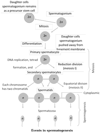
The process of formation of sperms in seminiferous tubules of the testis of the male animal is termed as spermatogenesis.
In mammals, testis have several coiled tubules in it called the seminiferous tubules. Sperms are formed in these tubules. The inner wall of seminiferous tubules is made up of germinal epithelium whose cells are cuboidal.
The endodermal cells of yolk sac migrate in testes and become primordial germ cells. Due to the division of these cells sperms are formed.
Some large cells are also found in this germinal epithelium. These are called the "Sertoli cells or Sustentacular cells". These cells provide nutrition to the maturing sperms in the form of Glycogen. For getting nutrition, the head of the sperms are submerged in the cytoplasm of sertoli cells.
Sertoli cells mainly provide nutrition and conserve the various stages of spermatogenesis. Spermatogenesis is a continuous process. To make it easier for study, it has been divided into the following steps -
(i) Formation of spermatid.
(ii) Spermiogenesis or Spermateleosis.
(i) Formation of spermatids : This process begins as the animal attains sexual maturity. The endodermal cells of the yolk sac which participate in this process are termed as the primordial germ cells. The process of formation of spermatids from primordial germ cells are termed as spermatocytosis. It has 3 sub-stages -
(a) Multiplication phase : During this process the primordial germ cells repeatedly undergo mitosis division, and as a result of these divisions spermatogonia are formed. Spermatogonia are diploid.
(b) Growth phase : Some spermatogonia either due to growth or due to food storage become 2 or 3 times of their original size, and are now known as primary spermatocytes. The remaining spermatogonia remain in the seminiferous tubules in the form of reserved stock. The primary - spermatocytes formed during the growth phase are diploid. Growth phase is the longest.
(c) Maturation phase : Primary - spermatocytes undergo Meiosis-I and as a result 2 haploid secondary spermatocytes are formed. This division is termed as First Maturation Division or Reductional division. Secondary spermatocytes undergo Meiosis II or equational division, and as result, 2 spermatids are formed from each secondary spermatocyte. Thus, from 1 diploid primary spermatocytes 2 secondary spermatocytes are formed on meiosis I and from 2 haploid secondary spermatocytes 4 spermatids are formed on meiosis-II. Metamorphosis of spermatids into sperms in known as Spermiogenesis or Spermatoliosis.
(ii) Spermatoliosis : The process of transformation of a round non-motile and haploid spermatid obtained from spermatocytosis into thread-like, motile and haploid sperm is termed as spermatoliosis. From different parts of the spermatid different parts of the sperm are formed. These are as follows -
(a) From nucleus and golgibody\[\to \]Head part
(b) From mitochondria\[\to \]Middle part
(c) The structure of the head of the sperm mainly depends on the structure of the nucleus. During spermatoliosis, nucleus contracts and acquires different shapes.
Structure of sperm
Structure of sperm has three parts
(1) Head (2) Middle piece (3) Tail
(1) Head : It is flat and oval in human sperm. It is composed of a large posterior nucleus and a small anterior acrosome.
Acrosome is formed from the golgi complex. It contains digestive enzyme hyaluronidase and proteinase. It is the capitis covering above the nucleus. It is surrounded by double membrane. Acrosome + its membrane are together called Galea-capatis. Acrosome plays important role in penetration of ovum by sperm.
Remaining part of the head is nucleus. Narrow space between the nucleus and the acrosome is termed as "perforatorium". Nucleus of the sperm is very small. In it nucleoplasm and nucleolus are absent. It contains only chromatin. At the base of the nucleus in a pit like depression proximal centriole is present. In between the head and the middle piece a small neck is present. In this neck part a distal centriole is located. Both the centrioles are at right angles to each other. Proximal centriole first induce cleavage in a fertilized egg. First spindle fibre forms from it. Distal centriole gives rise to the axial filament of the sperm. It has \[(9+2)\] microtubular arrangement.
(2) Middle piece : This is known as the energy-chamber of the sperm. Many mitochondria spirally surround the axonema, this is called "Nabenkern sheath". This part provides energy to the sperm for locomotion. In middle-piece, cytoplasm is found in the form of a thin-sheet called Manchett. In middle-part, axonema is surrounded by 9 solid fibres made up of proteins. At the posterior end of the middle-piece a Ring centriole is found. Its function is not known.
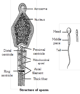
(3) Tail : The longest and the fibrous part of the sperm is termed its tail.
Sperm moves with the help of its tail. Basal granule of the tail is Distal centriole. Tail has 2 parts
(i) Main part : This part is broad. It contains cytoplasm and is surrounded by 2 solid fibres.
(ii) End piece : This part is narrow in it cytoplasm is absent only axonema is present. In it solid fibres are also absent. In the sperm of certain animals, tail is absent. e.g.,
(a) Ascaris : Tailless, ameboid sperms
(b) Cray fish : Tailless, stellate (star shape) sperms.
(c) Crab and lobser : Tailless sperms with 3 spines at apex.
(d) Biflagellage sperms : In Toad fish (Opsanus)
(e) In Opposum : Many sperms fuse together by their heads to form a "sperm-boat".
(f) Gastrapods have hexaflagellated sperms.
Oogenesis
Oogenesis takes place in the ovaries. Unlike sperm formation that starts at puberty, egg formation begins before birth but is completed only after fertilization. Oogenesis consists of three phases -
(a) Multiplication phase
During foetal development, endodermal cell of yolk sac enter into ovary and begins oogenesis.
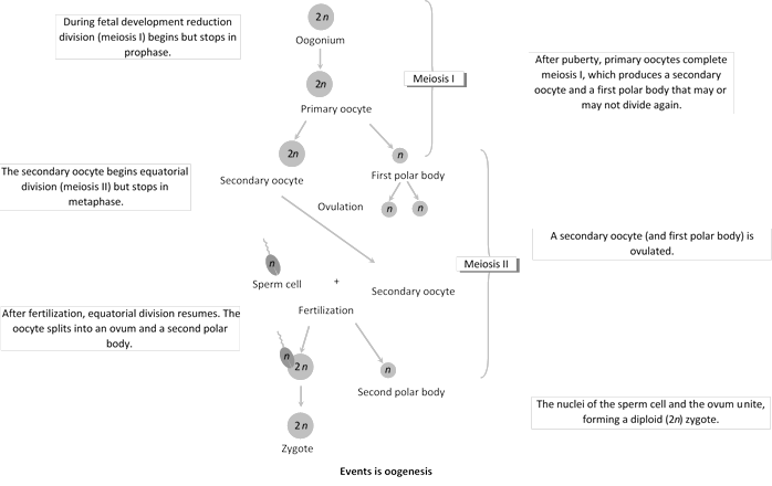
These cells undergo mitotic divisions, producing undifferentiated germ cells called oogonia or egg mother cells in the ovary. The oogonia have diploid, number of chromosome, 46 in humans. The oogonia multiply by mitotic divisions and produce ovigerous cords or egg tubes of pfluger in mammals.
(b) Growth phase : It is prolonged and slow. Oogonia form rounded masses or egg nests at the tips of egg tubes of pfluger.
An egg nest forms ovarian follicle (Graffian follicle) one central oogonium grows and functions as primary oocyte. The others form the covering follicular cells. the latter provide nourishment to primary oocyte. Some nourishment also comes from outside. Yolk is deposited in this state. This phenomenon is called vetellogenesis.
In cooperation with follicular cells, the enlarged primary oocyte secrete mucoprotein membrane or zona pellucida outside its own plasma membrane or vitelline membrane. There is increase in reserve food, size of nucleus, number of mitochondria; functioning of golgi apparatus and complexing of endoplasmic reticulam.
(c) Maturation phase : Meiosis occurs. Nucleus shifts towards animal pole and undergoes meiosis - I. A daughter nucleus alongwith small quantity of cytoplasm is extruded as primary polar body or polocyte below zona pellucida. Simultaneously primary oocyte is changed into haploid secondary oocyte. It proceeds with meiosis – II but stops at metaphase-II. Ovum is generally shed in secondary oocyte stage.
After fertilization, the second meiotic division is completed with unequal cytoplasmic cleavage. This forms a large cell the ootid with essentially whole of the cytoplasm, and a very small cell, the second polar body. The ootid and the second polar body are haploid as the second meiotic division is equational. The first polar body may divide at about the same time into two polar bodies. One primary oocyte forms, after two meiotic division, one haploid ootid and two or three haploid polar bodies. The ootid grows into a functional haploid ovum.
Structure of ovum
An ovum is generally spherical, nonmotile gamete with yolky cytoplasm and enclosed in one or more egg envelops. Size of ovum varies in different animals and depends upon the amount of yolk. Size of ovum varies from 10m to a few cm. Largest sized egg is of ostrich and is about \[170\times 135\,\,mm.\] Egg size and yolk amount are interdependent. It is about \[50\mu \] in many polychaete worms, \[150\mu \] in tunicates but very large sized in birds and reptiles. In mammals, it is generally microlecithal and about \[100\mu .\]
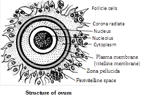
Human ovum is microlecithal with large amount of cytoplasm. Cytoplasm is differentiated into outer, smaller and transparent exoplasm or egg cortex and inner, larger and opaque endoplasm or ooplasm.
Egg envelopes. Human ovum is surrounded by a number of egg envelopes :
(a) Vitelline membrane : It is inner, thin, transparent and is secreted by ovum itself.
(b) Zona pellucida : It is middle, thick, transparent and non-cellular.
(c) Corona radiata : It is outer, thicker coat formed of radially elongated follicular cells. Between the vitelline membrane and zona pellucida, there is a narrow perivitelline space.
Differences between Spermatogenesis and Oogenesis
|
S.No. |
Characters |
Spermatogenesis |
Oogenesis |
|
1. |
Site of occurrence |
In the seminiferous tubules of testes. |
In the ovaries. |
|
2. |
Total period |
It is a continuous process and completed in 74 days in humans |
It is a discontinuous process and completed in a minimum 12-15 yrs. |
|
3. |
Growth phase |
Of shorter duration |
Of longer duration |
|
4. |
Yolk synthesis |
No yolk is synthesized in growth phase |
Vitellogenesis occurs in growth phase. |
|
5. |
Nuclear changes |
Nucleus becomes condensed by the loss of superfluous materials. |
Nucleus is bloated due to increase in necleoplasm. |
|
6. |
Number of gametes |
One spermatogonium forms 4 haploid sperms. |
One oogonium forms only one haploid ovum. |
|
7. |
Polar bodies |
Not formed. |
Two or three polar bodies are formed. |
|
8. |
Site of completion |
It is started and completed within the testes. |
It is started inside the ovary but is generally completed outside the ovary, into oviduct. |
|
9. |
Size of gametes formed |
Sperm is much smaller than spermatogonium. |
Ovum is much larger than oogonium. |
Types of eggs
(1) On the basis of amount and distribution of yolk
(i) Alecithal or Microlecithal or Oligolecithal or Meolecithal and Isolecithal or Homolecithal : The amount of yolk is very small in these types of eggs. (Oligolecithal or Microlecithal or Alecithal) and yolk is evenly distributed in these eggs (Isolecithal or Homolecithal). Examples - Egg of Amphioxus, Eutheria (Human egg), Metatheria and Sea-urchin.
(ii) Mesolecithal or Telolecithal eggs : In this type of egg the amount of yolk is moderate and yolk is concentrated in the basal part of egg (telolecithal egg). Examples - Egg of Amphibia, Petromyzon and Lung fishes.
(iii) Polylecithal or Macrolecithal or Megalecithal eggs : Eggs are with large amount of yolk e.g., eggs of shark, bony fish, Reptiles, birds, prototherian, concentrated mainly in vegetal pole.
In discoidal or highly telolecithal eggs, the yolk is enormous in amount and cytoplasm is confined to a disc like area on yolk. This disc of cytoplasm is called germinal disc. Example – Eggs of reptiles, birds, protherian mammals.
(iv) Centrolecithal : Yolk concentrate in centre e.g., Insects egg.
Smallest eggs are of 50m in the polychaeta and the largest eggs are of an ostrich.
(2) On the basis of fate
(i) Determinate / Mosaic eggs : Every part of fertilize egg has a definite fate, so that fate of every blastomere is determined from the beginning. It is found in invertebrates except echinoderms.
(ii) Indeterminate / Regulative eggs : The fate of different parts of egg or its blastomeres is not predetermined. Example - Echinoderms, Vertebrates.
(3) On the basis of shell
(i) Cleidoic eggs : Eggs surrounded by a hard shell are known as cleidoic eggs. These eggs are found in those animals which have a terrestrial mode of life of which lay eggs on land. These eggs have more amount of yolk. These are adaptations to terrestrial mode of life. Shell prevents the egg from dessication. e.g., - Eggs of "Reptiles". "Birds". "Insects" and "Prototherians".
(ii) Non - Cleidoic eggs : Eggs which are not surrounded by a hard shell are called Non-cleidoic eggs. These eggs are found in all oviperous animals which lay eggs in water and all viviperous animals. e.g., - All viviperous animals (Mammals) and all oviperous animals which lay eggs in water (Amphibians).
Classification of egg - membranes
On the basis of origin, egg-membranes are of 3 types -
(1) Primary egg membrane : This membrane is secreted by the egg (ovum) itself. e.g., - Vitelline membrane of human egg.
(2) Secondary egg membrane : This is found outside the primary egg membrane and is secreted by the ovary. e.g., Chorion of insect eggs, corona radiata and zona pellucida of human egg.
(3) Tertiary egg membrane : This present outside the primary and the secondary egg membrane. It is either secreted by the uterus or the oviduct. Egg jelly coat around frog's egg; albumen, shell membrane and shell of bird egg.
Functions of egg membranes
(1) To provide protection.
(2) To check polyspermy.
(3) To provide buoyancy to the amphinian eggs.
(4) To provide nutrition (Birds, Reptiles)
(5) To help in excretion (Allantois)
Different types of eggs
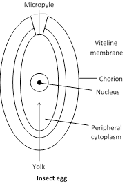
(1) Insect egg : Eggs of insects are megalecithal or polylecithal in them yolk is present in the centre, so the eggs are also centrolecithal. Eggs of insects are cigar like. Along with plasma-membrane the egg has 2 egg-membranes.
(i) Vitelline membrane : This is a primary egg membrane and the egg itself secretes it around.
(ii) Chorion : This is a secondary egg membrane and is secreted by the ovary. In Insect's egg tertiary egg-membranes is absent. Chorion of insect's egg is ornamented i.e. there are specific markings on its egg membrane which are characters of Taxonomic importance. In the egg, a hole termed as micropyle is present which is the port of entry for sperms. Its cytoplasm is divided into 2 parts -
(a) Central cytoplasm
(b) Peripheral cytoplasm
(a) Central cytoplasm : It is present in a very small amount in the centre of the egg. Egg nucleus is located in it.
(b) Peripheral cytoplasm : It is present in a very small amount along the periphery of the egg.
Yolk : In insect's egg yolk is present in a very large amount and this yolk is concentrated between the central and the Peripheral cytoplasm.
(2) Frog's egg : Eggs of frog are Telolecithal and Mesolecithal. The egg has 2 egg membranes.
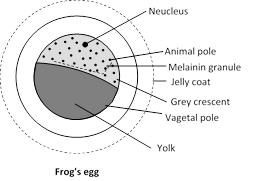
(i) Vitelline membrane : This is a primary membrane, secreted around by the egg itself.
(ii) Jelly – coat : This is a tertiary egg-membrane. It is secreted by the oviduct. Secondary egg-membrane are absent in these egg's. Internally, the egg is divided into 2 areas -
(a) Animal pole (b) Vegetal pole
(a) Animal pole : This part has more amount of cytoplasm in it and the egg nucleus is also located in it. In this part melanin granules are found which prevent the egg from harmful radiations. Due to these melanin granules the frog's egg is partly white and partly black. This helps in Camouflage. Sperm always enters inside the egg through the animal pole. The part from where the sperm enters inside the frog's egg in future forms the ventral part of the embryo. As the sperm enters inside the egg. The part directly opposite to the entry point becomes a clear-zone due to the rapid movement of melanin granules. this clear-zone is termed as the Grey-Crescent. This part with Grey-Crescent forms the dorsal part of the embryo in future.
(b) Vegetal pole : Here the yolk is concentrated in frog's egg, the part with cytoplasm in future forms the ectoderm. The Grey crescent part in future the Mesoderm and the part with yolk in future forms the endoderm.
Jelly-coats of all the eggs of a frog absorb water and swell up, to form a cluster of eggs termed as Spawn. Jelly-coat has air-bubbles, due to which the eggs don't drown. Jelly-coat is bitter in taste and so the eggs are protected from the enemies.


You need to login to perform this action.
You will be redirected in
3 sec

