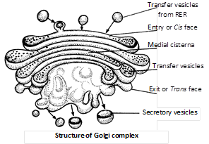
 Origin : Most accepted view is that golgi body originates from RER-that has lost its ribosomes from this RER arise transport vesicles that contain Golgi membrane and fuse with the saccule on the forming face of Golgi apparatus. This is why this face is called the forming face.
Functions
(1) The main function of golgi body is secretion, so it is large sized among the secretory cells.
(2) Glycosidation of lipids i.e., addition of oligosaccharides to produce glycolipids.
(3) Glycosylation of proteins i.e., addition of carbohydrate to produce glycoproteins.
(4) Formation of primary lysosomes.
(5) Golgi body forms the cell plate. During cell division by secreting hemicellulose formation of enzyme and hormones (Thyroxine) etc.
(6) In oocytes of animal, golgi apparatus functions as the centre around which more...
Origin : Most accepted view is that golgi body originates from RER-that has lost its ribosomes from this RER arise transport vesicles that contain Golgi membrane and fuse with the saccule on the forming face of Golgi apparatus. This is why this face is called the forming face.
Functions
(1) The main function of golgi body is secretion, so it is large sized among the secretory cells.
(2) Glycosidation of lipids i.e., addition of oligosaccharides to produce glycolipids.
(3) Glycosylation of proteins i.e., addition of carbohydrate to produce glycoproteins.
(4) Formation of primary lysosomes.
(5) Golgi body forms the cell plate. During cell division by secreting hemicellulose formation of enzyme and hormones (Thyroxine) etc.
(6) In oocytes of animal, golgi apparatus functions as the centre around which more... 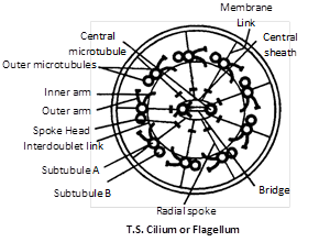 (1) Basal body : These are also termed as blepharoplast (kinetosome) or basal granule. It is present below the plasma membrane in cytoplasm. The structure is similar to centriole made of 9 triplets of microtubules.
(2) Rootlets : Made of microfilament and providing support to the basal body.
(3) Basal plate : Central fibril develop in this area. It is highly dense and lie above plasma-membrane. The basal body and the shaft at the level of plasma membrane.
(4) Shaft : It is the hair like projecting part of cilia and flagella which remains outside the cytoplasm. It has 9 doublet of microtubules in radial symmetry. These are called axonema. Each axonema has 11 fibrils, 9 in the periphery and 2 in the centre. The arrangement is called 9 + 2 pattern.
Chemical composition : Chemically, the central tubules are formed of dynein protein while the peripheral microtubules are formed of tubulin protein.
Type of flagella : There are two types of flagella.
(1) Tinsel type : In this, flagellum has lateral hair-like processes, called flimmers or mastigonemes.
(2) Whiplash type : In this, flagellum has no flimmers.
Functions
(1) They help in locomotion, respiration, cleaning, circulation, feeding, etc.
(2) Being protoplasmic structure they can function as sensory organs.
(3) They show sensitivity to changes in light, temperature and contact.
Difference between cilia and flagella
(1) Basal body : These are also termed as blepharoplast (kinetosome) or basal granule. It is present below the plasma membrane in cytoplasm. The structure is similar to centriole made of 9 triplets of microtubules.
(2) Rootlets : Made of microfilament and providing support to the basal body.
(3) Basal plate : Central fibril develop in this area. It is highly dense and lie above plasma-membrane. The basal body and the shaft at the level of plasma membrane.
(4) Shaft : It is the hair like projecting part of cilia and flagella which remains outside the cytoplasm. It has 9 doublet of microtubules in radial symmetry. These are called axonema. Each axonema has 11 fibrils, 9 in the periphery and 2 in the centre. The arrangement is called 9 + 2 pattern.
Chemical composition : Chemically, the central tubules are formed of dynein protein while the peripheral microtubules are formed of tubulin protein.
Type of flagella : There are two types of flagella.
(1) Tinsel type : In this, flagellum has lateral hair-like processes, called flimmers or mastigonemes.
(2) Whiplash type : In this, flagellum has no flimmers.
Functions
(1) They help in locomotion, respiration, cleaning, circulation, feeding, etc.
(2) Being protoplasmic structure they can function as sensory organs.
(3) They show sensitivity to changes in light, temperature and contact.
Difference between cilia and flagella
| Cilia | Flagella |
| More in number (may be upto 14,000 per cell). | Less in number (1-8). |
| Small sized \[(5-10\,\mu m).\] | Large sized (upto \[100-200\,\mu m).\] |
| more...
Discovery : Centrosome was first discovered by Van Benden (1887) and structure was given by T. Boweri.
Occurrence : It is found in all the animal cell except mature mammalian RBC’s. It is also found in most of protists and motile plant cells like antherozoids of ferns, zoospores of algae and motile algal forms e.g., Chlamydomonas but is absent in prokaryotes, fungi, gymnosperms and angiosperms.
Structure : Centrosome is without unit membrane structure. It is formed of two darkly stained granules called centrioles, which are collectively called diplosome. These centrioles are surrounded by a transparent cytoplasmic area called centrosphere of Kinetoplasm. Centriole and centrosphere are collectively called centrosome. Each centriole is a microtubular structure and is formed of microtubules arranged in 9+0 manner (all the 9 microtubules are peripheral in position). Inside the microtubules, there is an intra-centriolar or cart-wheel structure which is formed of a central hub (about \[25{AA}\] in diameter) and 9 radial spokes or radial fibres.
Chemical composition : Centrosome is lipoproteinaceous structure. The microtubules of centriole are composed of protein tubulin and some lipids. They are rich in ATPase enzyme.
Origin : The daughter centriole is formed from the pre-existing centriole in \[{{G}_{2}}\] of interphase so called self-replicating organelle.
Functions
(1) The centrioles help organising the spindle fibres and astral rays during cell division.
(2) They provide basal bodies which give rise to cilia and flagella.
Discovery : It was first discovered by Robert Hooke in 1665 in Cork. Cell wall is the outer most, rigid, protective, non living and supportive layer found in all the plant cells, bacteria, cyanobacteria and some protists. It is not found in animal cells.
Chemical composition : Mainly cell wall consists of two parts, matrix and cellulosic fibres (microfibrils). Matrix consists of hemicellulose, pectin, glycoproteins, lipids and water.In most of the plants cell wall is made up of cellulose
\[{{({{C}_{6}}{{H}_{10}}{{O}_{5}})}_{n}},\]
a polymer made-up of unbranched chain of glucose molecule linked by
\[\beta ,\,1-4\]
glycosidic bond. About 100 molecules of cellulose form a micelle, about 20 micelle form a microfibril and approx 200 microfibril form a fibril.
The cell wall of bacteria and the inner layer of blue green algae is made-up mucopeptide. It is a polymer of two amino sugars namely N-acetyl glucosamine (NAG) and N-acetyl muramic acid (NAM) held alternately in
\[\beta ,\,1-4-\]
linkage. In higher fungi, the cell wall is made up of chitin, polymer of glucosamine.
Structure : Cell wall consists of middle lamella, primary wall, secondary wall, tertiary wall.
(1) Middle lamella : Middle lamella is the outermost region which functions as a common cementing layer between two cells. It is absent on the outer free surface. It ruptures to create intercellular spaces. Middle lamella is formed of calcium and magnecium pectate. Fruit softening is due to gelatinisation of pectic compounds of middle lamella. Pectin is used as commercial jellying agent. Which is present outside the primary wall.
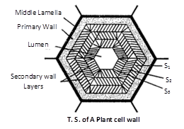 (2) Primary wall : A young plant cell forms a single layer of wall material. This layer is known as the primary cell wall. The primary wall is thin, elastic and capable of expansion in a growing cell. It grows by intussusception. Meristematic and parenchymatous cells have primary cell wall only. The cells of leaves and fruits too have only primary wall. It has more hemicellulose and less cellulose.
(3) Secondary wall : In mature cell, more layers of wall material are added internal to the primary wall. These are called the secondary cell wall. Growth by addition of new wall material on the primary wall is called accretion. It has more cellulose and less hemicellulose. The secondary wall is thick and rigid. It usually consists of three layers, which are often named
\[{{S}_{1}},{{S}_{2}}\,\text{and }{{\text{S}}_{\text{3}}}.\]
It is found in collenchyma and sclerenchyma cells, xylem vesseles.
(4) Tertiary wall : Sometimes tertiary wall is laid down on secondary wall, e.g., tracheids of gymnosperms. It is composed of cellulose and xylan.
Origin : A cell wall is originate at telophase stage of cell division. The plane and place of cell wall is determined by the microtubules. Fragments of ER and vesicles of golgi body alligned at the equator, called as phragmoplast, later which forms the cell plate. The synthesis of cellulose takes place by the help of enzyme cellulose synthase present in the plasma membrane. The more...
(2) Primary wall : A young plant cell forms a single layer of wall material. This layer is known as the primary cell wall. The primary wall is thin, elastic and capable of expansion in a growing cell. It grows by intussusception. Meristematic and parenchymatous cells have primary cell wall only. The cells of leaves and fruits too have only primary wall. It has more hemicellulose and less cellulose.
(3) Secondary wall : In mature cell, more layers of wall material are added internal to the primary wall. These are called the secondary cell wall. Growth by addition of new wall material on the primary wall is called accretion. It has more cellulose and less hemicellulose. The secondary wall is thick and rigid. It usually consists of three layers, which are often named
\[{{S}_{1}},{{S}_{2}}\,\text{and }{{\text{S}}_{\text{3}}}.\]
It is found in collenchyma and sclerenchyma cells, xylem vesseles.
(4) Tertiary wall : Sometimes tertiary wall is laid down on secondary wall, e.g., tracheids of gymnosperms. It is composed of cellulose and xylan.
Origin : A cell wall is originate at telophase stage of cell division. The plane and place of cell wall is determined by the microtubules. Fragments of ER and vesicles of golgi body alligned at the equator, called as phragmoplast, later which forms the cell plate. The synthesis of cellulose takes place by the help of enzyme cellulose synthase present in the plasma membrane. The more...
Cytology : (Gk Kyios = cell ; logas = study) It is the branch of biology. Which comprises the study of cell structure and function. “Cell is the structural and functional unit of all living beings”. Study of metabolic aspects of cell components is called cell biology.
Robert Hooke (1665) discovered hollow cavities (empty boxes) like compartments in a very thin slice of cork (cell wall) under his microscope. He wrote a book “Micrographia” and coined the term cellula, which was later changed into cell. Grew and Malpighi also observed small structures in slice of plants and animals. Leeuwenhoek was the first to see free cells and called them “wild animalcules” and published a book “The secret of nature”. He observed bacteria, protozoa, RBCs, sperms, etc. under his microscope.
Cell theory : H.J. Dutrochet (1824) a French worker gave the idea of cell theory.
The actual credit for cell theory goes to two German scientists, a Botanist M.J. Schleiden (1838) and a Zoologist T. Schwann (1839). They gave the concept “all living organisms are composed of cell”. Schleiden and Schwann both supported the theory of “spontaneous generation”. They also mentioned that “the new cell arises from nucleus by budding”.
Exceptions to the cell theory : Viruses, viroids and prions are an exception to the cell theory as they are obligate parasites (sub–cellular in nature).
Modification of cell theory : Modification of cell theory was done by Rudolf Virchow (1855). He proposed the “law of cell lineage” which states that cell originates from pre-existing cells. i.e., (omnis cellula-e-cellula). It is also called “cell principle” or “cell doctrine”. It states :
(1) Life exists only in cells.
(2) Membrane bound cell organelles of the protoplasm do not survive alone or outside the protoplasm.
(3) Cells never arise de novo. The new cells are like the parent cell in all respect.
(4) All cells have similar fundamental structure and metabolic reactions.
(5) Cells display homeostasis and remain alive.
(6) Genetic information is stored in DNA and expressed within the cells.
(7) DNA controls structure and working of a cell.
The cell as a self contained unit : Autonomy of a cell is believed due to presence of DNA and its expressibility, otherwise, cell components have different shape and function. It has two positions.
(1) Autonomy in unicellular organisms : Unicellular organisms leads to a totally independent life due to different shape, size and role of different organelles shows division of labour. All these display homeostasis. Unicellular organisms are more active due to large surface volume ratio.
(2) Autonomy in multicellular organisms : In multicellular organisms life activities are displayed by each of the cells independently. Multicellular organisms have one thing advantage over unicellular organisms is division of labour.
Cellular totipotency : Totipotency was suggested by Haberlandt (1902). When cells have tendency or ability to divide and redivide the condition of the cell is called totipotent and this phenomenon is called totipotency. Steward et.al. showed the phenomenon of cellular totipotency in carrot culture.
Surface more...
Mitochondria (Gk. Mito = thread ; chondrion = granule) are semi autonomous having hollow sac like structures present in all eukaryotes except mature RBCs of mammals and sieve tubes of phloem. Mesosomes of prokaryotes (bacteria) is analogous to mitochondrion in eukaryotes.
Mitochondria are also called chondriosome, chondrioplast, plasmosomes, plastosomes and plastochondriane.
Discoveries
(1) These were first observed in striated muscles (Voluntary) of insects as granules by Kolliker (1880), he called them “sarcosomes”.
(2) Flemming (1882) called them “fila” for thread like structure.
(3) Altman (1890) called them “bioplast”.
(4) C. Benda (1897) gave the term mitochondria.
(5) F. Meves (1904) observed mitochondria in plant (Nymphaea).
(6) Michaelis (1898) demonstrated that mitochondria play a significant role in respiration.
(7) Bensley and Hoerr (1934) isolated mitochondria from liver cells.
(8) Seekevitz called them “Power house of the cell”.
(9) Nass and Afzelius (1965) observed first DNA in mitochondria.
Number of mitochondria : Presence of mitochondria depends upon the metabolic activity of the cell. Higher is the metabolic activity, higher is the number e.g., in germinating seeds.
(1) Minimum number of mitochondria is one in Microasterias, Trypanosoma, Chlorella, Chlamydomonas (green alga) and Micromonas. Maximum numbers are found (up to 500000) in flight muscle cell, (up to 50000) in giant Amoeba called Chaos – Chaos. These are 25 in human sperm, 300 – 400 in kidney cells and 1000 – 1600 in liver cells.
(2) Mitochondria of a cell are collectively called chondriome.
Size of mitochondria : Average size is \[0.51.00\,\,\mu \,m\] and length up to \[110\,\,\mu \,m.\] Smallest sized mitochondria in yeast cells \[(1\,\mu \,{{m}^{3}}).\] and largest sized are found in oocytes of Rana pipiens and are \[2040\,\,\mu \,m.\]
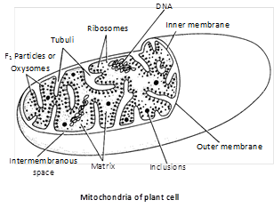
Ultrastructure : Mitochondrion is bounded by two unit membranes separated by perimitochondrial space (6 – 10nm wide). The outer membrane is specially permeable because of presence of integral proteins called porins. The inner membrane is selective permeable. The inner membrane is folded or convoluted to form mitochondrial crests. In animals these are called cristae and in plants these folding are called tubuli or microvili.
The matrix facing face is called ‘M’ face and face towards perimitochondrial space is called ‘C’ face. The ‘M’ face have some small stalked particles called oxysomes or \[{{F}_{1}}\] particle or elementory particle or Fernandez – Moran Particles (\[{{10}^{4}}{{10}^{5}}\] per mitochondria). Each particle is made up of base, stalk and head and is about 10nm in length.
Oxysomes have ATPase enzyme molecule (Packer, 1967) and therefore, responsible for ATP synthesis. These elementary particles are also called \[{{F}_{0}}\text{ }{{F}_{1}}\] particles. The \[{{F}_{1}}\] particle is made up of five types of subunits namely \[\alpha ,\,\beta ,\,\gamma ,\,\delta \] and \[\varepsilon .\] of these \[\alpha \] is heaviest and \[\varepsilon \] is lightest. \[{{F}_{0}}\] particles synthesize all the enzymes required to operate Kreb’s cycle.
 Semi-autonomous nature of mitochondrion : Mitochondria contain all requirements of protein synthesis :
(1) 70 S ribosomes.
(2) DNA molecules more...
Semi-autonomous nature of mitochondrion : Mitochondria contain all requirements of protein synthesis :
(1) 70 S ribosomes.
(2) DNA molecules more...
It is a technique of studying the route of chemicals in chemical reactions taking place inside the cell and organisms with the help of radioactive isotope. e.g., \[^{14}C,{{\,}^{3}}H,{{\,}^{32}}P.\]
In this technique the radioisotopes are incorporated into the precursor molecule. Then the labelled precursor molecules introduced into the cells and their path is followed with the help of their radiations.
Radioactive precursors emit radiations and their position in the cell is located by bringing the cell in contact with a photographic plate or film.
\[^{32}P\]and \[^{14}C\] are used for the study of nucleic acids and photosynthesis (Melvin Calvin) respectively.
Current Affairs CategoriesArchive
Trending Current Affairs


You need to login to perform this action. |

