Category : 11th Class
It is the process by which a mature cell divides and forms two nearly equal daughter cells which resemble the parental cell in a number of characters.
In unicellular organisms, cell division is the means of reproduction by which the mother cell produces two or more new cells. In multicellular organism also, new individual develop from a single cell. Cell division is central to life of all cell and is essential for the perpetuation of the species.
Discovery : Prevost and Dumans (1824) first to study cell division during the cleavage of zygote of frog. Nagelli (1846) first to propose that new cells are formed by the division of pre-existing cells.
Rudolf Virchow (1859) proposed “omnis cellula e cellula” and “cell lineage theory”.
A cell divides when it has grown to a certain maximum size which disturb the karyoplasmic index (KI)/Nucleoplasmic ratio (NP)/Kernplasm connection.
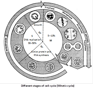
Cell cycle : Howard and Pelc (1953) first time described it. The sequence of events which occur during cell growth and cell division are collectively called cell cycle. Cell cycle completes in two steps:
(1) Interphase, (2) M-phase/Dividing phase
(1) Interphase : It is the period between the end of one cell division to the beginning of next cell division. It is also called resting phase or not dividing phase. But, it is actually highly metabolic active phase, in which cell prepares itself for next cell division. In case of human beings it will take approx 25 hours. Interphase is completed in to three successive stages.
G1 phase/Post mitotic/Pre-DNA synthetic phase/gap Ist : In which following events take place.
(i) Intensive cellular synthesis.
(ii) Synthesis of rRNA, mRNA ribosomes and proteins.
(iii) Metabolic rate is high.
(iv) Cells become differentiated.
(v) Synthesis of enzymes and ATP storage.
(vi) Cell size increases.
(vii) Decision for a division in a cell occurs.
(viii) Substances of G stimulates the onset of next S – phase.
(ix) Synthesis of NHC protein, carbohydrates, proteins, lipids.
(x) Longest and most variable phase.
(xi) Synthesis of enzyme, amino acids, nucleotides etc. but there is no change in DNA amount.
S-phase/Synthetic phase
(i) DNA replicates and its amount becomes double \[(2C-4C).\]
(ii) Synthesis of histone proteins and NHC (non-histone chromosomal proteins).
(iii) Euchromatin replicates earlier than heterochromatin.
(iv) Each chromosome has 2 chromatids.
G2-phase/Pre mitotic/Post synthetic phase/gap-IInd
(i) Mitotic spindle protein (tubulin) synthesis begins.
(ii) Chromosome condensation factor appears.
(iii) Synthesis of 3 types of RNA, NHC proteins, and ATP mole.
(iv) Duplication of mitochondria, plastids and other cellular macromolecular complements.
(v) Damaged DNA repair occur.
(2) M-phase/Dividing phase/Mitotic phase : It is divided in to two phases, karyokinesis and cytokinesis.
Duration of cell cycle : Time period for \[{{G}_{1}},S,{{G}_{2}}\] and M-phase is species specific under specific environmental conditions. e.g., 20 minutes for bacterial cell, 8-10 hours for intestinal epithelial cell, and onion root tip cells may take 20 hours.
\[{{\mathbf{G}}_{\mathbf{0}}}\mathbf{-}\]phase (Lajtha, 1963) : The cells, which are not to divide further, do not proceed beyond the \[{{G}_{1}}\] phase and start undergoing differentiation into specific type. Such cells are said to be in \[{{G}_{0}}\] phase.
Types of cell division : It is of three types, Amitosis, Mitosis and Meiosis.
![]()
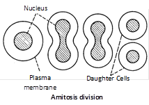
Amitosis (Gk. Amitos = without thread; osis = state). It is also called as direct cell division. It was discovered by Remak (1855) in RBC of chick embryo. In this division there is no differentiation of chromosomes and spindle. The nuclear envelope does not degenerate. The nucleus elongates and constricts in the middle to form two daughter nuclei. This is followed by a centripetal constriction of the cytoplasm to form two daughter cells. It is primitive type of division occuring in prokaryotes, protozoans, yeasts, foetal membrane of mammals, cartilage of mammals etc.
![]()
Mitosis (Gk. Mitos = thread; osis = state). It is also called indirect cell division or somatic cell division or equational division. In this, mature somatic cell divides in such a way that chromosomes number is kept constant in daughter cells equal to those in parent cell. So it is called equational division.
Discovery : Mitosis was first observed by Strasburger (1875) and in animal cell by W.flemming (1879) term mitosis was given by Flemming (1882).
Occurrence : Mitosis is the common method of cell division. It takes place in the somatic cells in the animals and plants. Hence, it is also known as the somatic division. In plants mitosis occurs in the meristematic cells e.g., root apex and shoot apex.
Process of mitosis : Mitosis is completed in two steps.
Karyokinesis : (Gk. Karyon = nucleus; kinesis = movement) Division of nucleus. Term given by Schneider (1887). Karyokinesis it takes \[5-10%\] (shortest phase) time of whole division. It comprises four phases i.e., Prophase, Metaphase, Anaphase, Telophase.
(1) Prophase : It is longest phase of karyokinesis.
(i) Chromatin fibres thicken and shorter to form chromosomes which may overlap each other and appears like a ball of wool. i.e., Spireme stage.
(ii) Each chromosome divides longitudinally into 2 chromatids which remain attached to centromere.
(iii) Nuclear membrane starts disintegrating except in dinoflagellates.
(iv) Nucleolus starts disintegrating.
(v) Cells become viscous, refractive and oval in outline.
(vi) Spindle formation begins.
(vii) Cell cytoskeleton, golgi complex, ER, etc. disappear.
(viii) In animal cells, centrioles move towards opposite sides.
(ix) Lampbrush chromosomes can be studied well.
(x) Small globular structure (beaded) on the chromosome are called chromomeres.
(xi) Spindle is formed from centriole (in animal cells) or MTOC (microtubule organising centre) in plant cells successively called astral and anastral spindle.
(2) Metaphase
(i) Chromosomes become maximally distinct i.e., size can be measured.
(ii) A colourless, fibrous, bipolar spindle appears.
(iii) Spindle fibre are made up of 97% tubulin protein and 3% RNA.
(iv) Chromosomes move towards equatorial plane of spindles called congression and become arranged with their arms directed towards pole and centromere towards equator.
(v) Spindle fibres attach to kinetochores.
(vi) Metaphase is the best stage for studying chromosome morphology (structure, size, number).
(vii) Spindle has two type of fibres
(a) Continous fibre (run from pole to pole).
(b) Discontinous fibre (between pole to centromeres).
(3) Anaphase
(i) Centromere splits from the middle and two chromatids gets separated.
(ii) Both the chromatids move towards opposite poles due to repulsive force called anaphasic movement.
(iii) Anaphasic movement is brought about by the repolymerisation of continuous fibres and depolymerisation of chromosomal fibres. Formation and expansion of interzonal fibres.
(iv) Different shape of chromosomes (V, J, I or L shapes) become evident during chromosome movement viz. metacentric acrocentric etc.
(v) The centromere faces towards equator.
(vi) The chromatids are moved towards the pole at a speed of \[1\,\mu \,m\]/minute. About 30 ATP molecules are used to move one chromosome from equator to pole.
(vii) Shape of chromosome is best studied at anaphase.
(4) Telophase
(i) Chromosomes reached on poles by the spindle fibers and form two groups.
(ii) Chromosomes begin to uncoil and form chromatin net.
(iii) The nuclear membrane and nucleolus reappear.
(iv) Two daughter nuclei are formed.
(v) Golgi complex and ER etc., reform.
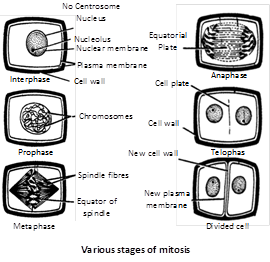
Cytokinesis : (\[Gk\]kitos = cell; kinesis = movement) Division of cytoplasm, Term given by Whiteman (1887). Division of cytoplasm into 2 equal parts. In animal cell, it takes place by cell furrow method and in plant cells by cell plate method.

Significance of mitosis
(1) It keeps the chromosome number constant and genetic stability in daughter cells, so the linear heredity of an organism is maintained. All the cells are with similar genetic constituents.
(2) It provides new cells for repair and regeneration of lost parts and healing of the wounds.
(3) It helps in asexual reproduction by fragmentation, budding, stem cutting, etc.
(4) Somatic variations when maintained by vegetative propagation can play important role in speciation.
(1) Intranuclear or Promitosis : In this nuclear membrane is not lost and spindle is formed inside the nuclear membrane e.g., Protozoans (Amoeba) and yeast. It is so as centriole is present within the nucleus.
(2) Extranuclear or Eumitosis : In this nuclear membrane is lost and spindle is formed outside nuclear membrane e.g., in plants and animals.
(3) Endomitosis : Chromosomes and their DNA duplicate but fail to separate which lead to polyploidy e.g., in liver of man, both diploid (2N) and polyploid cells (4N) have been reported. It is also called endoduplication and endopolyploidy.
(4) Dinomitosis : In which nuclear envelope persists and microtubular spindle is not formed. During movement the chromosomes are attached with nuclear membrane.
Mitotic poision : The agents which inhibit cell division.
(1) Azides and Cyanides : Inhibit prophase.
(2) Colchicine : Inhibits spindle formation at metaphase.
(3) Mustard gas : Agglutinates the chromosomes.
(4) Chalones : These were first reported by Laurence and Bullough (1960). They are peptides and glycoproteins secreated by extracellular fluid of healthy cells and inhibit cellular division.
Karyochoriosis : A type of mitosis in fungi in which is intranuclear nucleus divides by furrow formation.
Difference between animal and plant cells (Mitosis)
|
Animal cells |
Plant cells |
|
Centrioles present at spindle poles. |
Centrioles lacking at spindle poles. |
|
Asters are formed (amphiastral). |
No asters are formed (anastral). |
|
Cytokinesis by furrowing of cytoplasm. |
Cytokinesis mostly by cell plate formation. |
|
Furrow extends centripetally |
Cell plate grows centrifugally. |
|
Microfilament ring brings about cleavage. |
Microfilaments have no role in cytokinesis. |
|
Occurs nearly in all tissues. |
Occurs mainly at meristems. |
|
Cell becomes rounded and its cytoplasm more viscous at the time of mitosis. |
Cell does not change form or nature at the time of mitosis. |
|
Midbody is formed at the equator of the spindle. |
Equator of the spindle changes into phragmoplast. |
|
Intercellular spaces appear between the daughter cells. |
Daughter cells remain adhered together by middle lamella. |
|
Animal mitosis is controlled by certain mitogens. |
Plant mitosis is usually controlled by a hormone cytokinin. |
![]()
Meiosis (Gr. meio – to lessen, osis – state). Meiosis is a much slower process than mitosis. It is a double division that occurs in a mature diploid reproductive cell (2x) in which nucleus divides twice but chromosome (DNA) replicates only once to form four haploid cells, each having the half the number of chromosomes present in the parent cell. As it causes reduction in the number of chromosomes, it is known as reduction division. Meiosis in a cell occurs only once. The so formed haploid cells do not further undergo meiosis because there is no synaptinemal complex in haploid genome.
Discovery : It was first demonstrated by Van Beneden (1887) but was described by Winiwarter (1900). Term “meiosis” was given by Farmer and Moore (1905). Gregoire used the term meiosis I and II.
Occurrence : It is found in special types and at specific period. It is reported in diploid germ cells of sex organs (e.g., primary spermatocytes of testes to form male gametes called spermotozoa and primary oocytes to form female gametes called ova in animals) and in pollen mother cells (microsporocytes) of anther and megasporocyte of ovule of ovary of flowers in plant to form the haploid spores. The study of meiosis in plants can be done in young flower buds.
Process of meiosis : Meiosis is completed in two steps, meiosis I and meiosis II
Meiosis-I : In which the actual chromosome number is reduced to half. Therefore, meiosis I is also known as reductional division or heterotypic division. It results in the formation of two haploid cells from one diploid cell. It is divided into two parts, karyokinesis I and cytokinesis I.
Karyokinesis-I : It involves division of nucleus. It is divided into four phases i.e., prophase, metaphase, anaphase, telophase.
(1) Prophase-I : It is of longest phase of karyokinesis of meiosis. It is again divisible into five subphases i.e., leptotene, zygotene, pachytene, diplotene and diakinesis.
(i) Leptotene/Leptonema
(a) Chromosomes are long thread like with chromomeres on it.
(b) Volume of nucleus increases.
(c) Chromatin network has half chromosomes from male and half from female parent.
(d) Chromosome with similar structure are known as homologous chromosomes.
(e) Leptonemal chromosomes have a definite polarization and forms loops whose ends are attached to the nuclear envelope at points near the centrioles, contained within an aster. Such peculiar arrangement is termed as bouquet stage (in animals) and syndet knot (in plants).
(g) Lampbrush chromosome found in oocyte of amphibians is seen in leptotene.
(ii) Zygotene/Zygonema
(a) Pairing or “synapsis” of homologous chromosomes takes place in this stage.
(c) Paired chromosomes are called bivalents, which by furthur molecular packing and spiralization becomes shorter and thicker.
(d) Pairing of homologous chromosomes in a zipper-fashion. Number of bivalents (paired homologous chromosomes) is half to total number of chromosomes in a diploid cell. Each bivalent is formed of one paternal and one maternal chromosome (i.e., one chromosome derived from each parent).
(e) Under EM, a filamentous ladder like nucleoproteinous complex, called synaptinemal. Synaptinemal complex is seen between the homologous chromosomes which was discovered by “Moses” (1953).
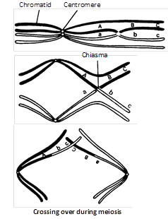
(iii) Pachytene/Pachynema
(a) In the tetrad, two similar chromatids of the same chromosome are called sister chromatids and those of two homologous chromosomes are termed non-sister chromatids.
(b) Crossing over i.e., exchange of segments between non-sister chromatids of homologous chromosome occurs at this stage.
It takes place by breakage and reunion of chromatis segments. Breakage called nicking, is assisted by an enzyme endonuclease and reunion termed annealing is added by an enzyme ligase. Breakage and reunion hypothesis proposed by Darlington (1937).
(c) Chromatids of pachytene chromosome are attached with centromere.
(d) A tetrad consists of two sets of homologous chromosomes each with two chromatids. Each tetrad has four kinetochore (two sister and two homologous).
(e) A number of electron dense bodies about 100 nm in diameter are seen at irregular intervals within the centre of the synaptonemal complex, known as recombination nodules.
(f) DNA polymerase is responsible for the repair synthesis.
(iv) Diplotene/Diplonema
(a) At this stage the paired chromosomes begin to separate (desynapsis).
(b) Cross is formed at the place of crossing over between non-sister chromatids.
(c) Homologous chromosomes move apart they remain attached to one another at specific points called chiasmata.
(d) At least one chiasma is formed in each bivalent.
(e) Chromosomes are attached only at the place of chiasmata.
(f) Chromatin bridges are formed in place of synaptonemal complex on chiasmata.
(g) This stage remains as such for long time.
(v) Diakinesis
(a) Chiasmata moves towards the ends of chromosomes. This is called terminalization.
(b) Chromatids remain attached at the place of chiasma only.
(c) Nuclear membrane and nucleolus degenerates.
(d) Chromosome recondense and tetrad moves to the metaphase plate.
(e) Formation of spindle.
(f) Bivalents are irregularly and freely scattered in the nucleocytoplasmic matrix.
When the diakinesis of prophase-I is completed than cell enters into the metaphase-I.

(2) Metaphase-I
(i) Chromosome come on the equator.
(ii) Bivalents arrange themselves in two parallel equatorial or metaphase plates. Each equatorial plate has one genome.
(iii) Centromeres of homologous chromosomes lie equisdistant from equator and are directed towards the poles while arms generally lie horizontally on the equator.
(iv) Each homologous chromosome has two kinetochores and both the kinetochores of a chromosome are joined to the chromosomal or tractile fibre of same side.
(3) Anaphase-I
(i) It involves separartion of homologous chromosomes which start moving opposite poles so each tetrad is divided into two daughter dyads. So anaphase-I involves the reduction of chromosome number, this is called disjunction.
(ii) The shape of separating chromosomes may be rod or J or V-shape depending upon the position of centromere.
(iii) Segregation of mendalian factors or independent assortment of chromosomes take place. In which the paternal and maternal chromosomes of each homologous pair segregate during anaphase-I which introduces genetic variability.
(4) Telophase-I
(i) Two daughter nuclei are formed but the chromosome number is half than the chromosome number of mother cell.
(ii) Nuclear membrane reappears.
(iii) After telophase I cytokinesis may or may not occur.
(iv) At the end of Meiosis I either two daughter cells will be formed or a cell may have two daughter nuclei.
(v) Meiosis I is also termed as reduction division.
(vi) After meiosis I, the cells in animals are reformed as secondary spermatocytes or secondary oocytes; with haploid number of chromosomes but diploid amount of DNA.
(vii) Chromosomes undergo decondensation by hydration and despiralization and change into long and thread like chromation fibres.
Interphase : Generally there is no interphase between meiosis-I and meiosis-II. A brief interphase called interkinesis, or intrameiotic interphase. There is no replication chromosomes, during this interphase.
Cytokinesis-I : It may or may not be present. When present, it occurs by cell-furrow formation in animal cells and cell plate formation in plant cells.
Significance of meiosis-I
(1) It separates the homologous chromosomes to reduce the chromosome number to the haploid state, a necessity for sexual reproduction.
(2) It introduces variation by forming new gene combinations through crossing over and random assortment of paternal and maternal chromosomes.
(3) It induces the cells to produce gametes for sexual reproduction or spores for asexual reproduction.
Meiosis-II : It is also called equational or homotypical division because the number of chromosomes remains same as after meiosis-I. It is of shorter duration than even typical mitotic division. It is also divisible into two parts, Karyokinesis-II and Cytokinesis-II.
Karyokinesis-II : It involves the separation of two chromatids of each chromosome and their movement to separate cells. It is divided in four phases i.e., Prophase-II, Metaphase-II. Anaphase-II and Telophase-II.
Almost all the changes of Karyokinesis-II resembles to mitosis which involves.
(1) It starts just after end of telophase I.
(2) Each daughter cell (nucleus) undergoes mitotic division.
(3) It is exactly similar to mitosis.
(4) At the end of process, cytokinesis takes place.
(5) Four daughter cells are formed after completion.
(6) The sister kinetochores of one chromosome are separated.
(7) The four daughter cells receive one chromatid each of the tetravalent.
(8) Centromere divide at anaphase II.
(9) Spindle fibres contract at prophase II.
Cytokinesis-II : It is always present and occurs by cell furrow formation in animal cell and cell plate formation in plant cell.
So by meiosis, a diploid parental cell divides twice forming four haploid gametes or sex cells, each having half the DNA amount than that of the parental cell and one-fourth of DNA present in the cell at the time of beginning of meiosis.
Significance of meiosis-II
(1) Constancy of chromosome number in successive generation is brought by process.
(2) Chromosome number becomes half during meiosis.
(3) It helps in introducing variations and mutation.
(4) It brings about gamete formation.
(5) It maintains the amount of genetic informative material.
(6) Sexual reproduction includes one meiosis and fusion.
(7) The four daughter cells will have different types of chromatids.
Types of meiosis
(1) Gametic/Terminal meiosis : In many protozoans, all animals and some lower plants, meiosis takes place before fertilization during the formation of gametes. Such a meiosis is described as gametic or terminal.
(2) Zygotic or Initial meiosis : In fungi, certain protozoan groups, and some algae fertilization is immediately followed by meiosis in the zygote, and the resulting adult organisms are haploid. Such a meiosis is said to be zygotic or initial. This type of life cycle with haploid adult and zygotic meiosis is termed the haplontic cycle.
(3) Sporogenetic / Intermediate meiosis
(i) Diploid sporocytes or spore mother cells of sporophytic plant, undergo meiosis to form the haploid spores in the sporangia.
(ii) Haploid spore germinates to form haploid gametophyte which produces the haploid gametes by mitosis.
(iii) Haploid gametes fuse to form diploid zygote which develops into diploid sporophyte by mitotic divisions. e.g., In higher plants like pteridophytes, gymnosperms and angiosperms.


You need to login to perform this action.
You will be redirected in
3 sec

