Blood Circulation In Human
Category : 11th Class
The physiology of blood circulation was first described by Sir William Harvey in 1628. The blood circulation in our body is divisible into 3 circuits -
(1) Coronary circulation : It involves blood supply to the heart wall and also drainage of the heart wall.
(i) Coronary arteries : One pair, arising from the aortic arch just above the semilunar valves. They break up into capillaries to supply oxygenated blood to the heart wall.
(ii) Coronary veins : Numerous, collecting deoxygenated blood from the heart wall and drains it into right auricle through coronary sinus which is formed by joining of most of the coronary veins. But some very fine coronary veins, called venae cordis minimae open directly in the right auricle by small sized openings called foramina of Thebesius.
(2) Pulmonary circulation : It includes circulation between heart and lungs. The right ventricle pumps deoxygenated blood into a single, thick vessel called pulmonary aorta which ascends upward and outside heart gets divided into longer, right and shorter, left pulmonary arteries running to the respective lungs where oxygenation of blood takes place. The oxygenated blood from lungs is returned to the left auricle by four pulmonary veins. Left auricle pumps this blood into the left ventricle.
(3) Systemic circulation : In this, circulation of blood occurs between heart and body organs. The left ventricle pumps the oxygenated blood into systemic arch which supplies it to the body organs other than lungs through a number of arteries. The deoxygenated blood from these organs is returned to the right auricle through two large veins (precaval and post caval). Right auricle pumps this blood into the right ventricle. Thus, the sytemic circulation involves two circuits -
Arterial system
It involves aorta, arteries, arterioles and meta-arterioles. It supplies oxygenated blood to all parts of the body except lungs.
The left ventricle of the heart pumps the oxygenated blood into a single, question marked shaped, long vessel called left carotid-systemic aorta. It is the largest blood vessel of the body.
After ascending from the heart, the systemic aotra turns and descends down to the level of lower border of fourth lumbar vertebra. At its distal extremity, it bifurcates into right and left common iliac arteries. The sytemic aorta has following parts -
(1) Ascending aorta : It gives off left and right coronary arteries.
Brachiocephalic (innominate) : Unpaired, largest branch of the aorta divides into right subclavian towards right side and right common carotid towards left side. Right subclavian gives off vertebral artery (supplies to head and part of right shoulder) and then enters into right arm, now called axillary artery or brachial artery, which divides into ulnar and radial arteries in the region of elbow. The right common carotid, enters into head and divides into external and internal carotids which supply the right parts of head by their branches.
Left common carotid : Unpaired artery, enters into head and divides into left external and internal carotids which supply the left parts of the head by their tributaries. The external carotids of both sides provide blood to thyroid gland, tongue, throat, face, ear, scalp. The internal carotids of both sides supply to brain, eye, inner part of nose and forehead. These internal carotids go upward and enter skull through foramen magnum and unite at the base of brain along with the vertebral arteries of both sides. So, there is formation of a ring shaped artery called as “Circle of willis”. From this circle, many branches or arteries arise which go to different parts of brain.
In frog, the internal carotid has at its base, carotid labyrinth (spongy mass of non-contractile fibro-elastic tissue) which acts as a sensory organ to detect blood pressure in artery.
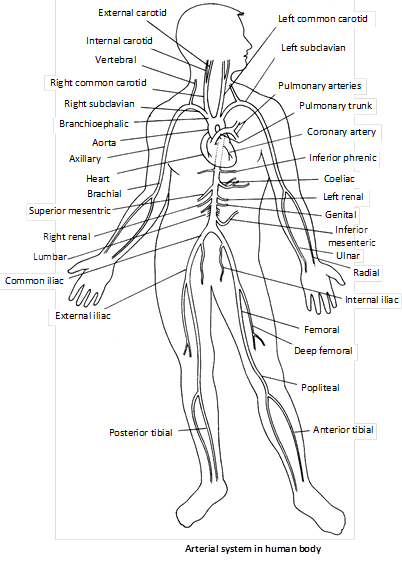
Left subclavian artery : Unpaired artery, it gives off a left vertebral artery (supplies to head and part of left shoulder) and then enters into left arm, now called left axillary artery or left brachial artery which divides into ulnar and radial arteries in the region of elbow.
(2) Descending aorta : The aorta turns towards the back of heart and finally converts into dorsal aorta. The descending dorsal aorta is called thoracic aorta in throcic region and abdominal aorta in abdominal region.
From thoracic segment of aorta : Several pairs of small arteries arise in this region to supply various parts such as pericardium (pericardial artery); lungs and bronchi (bronchial artery); oesophagus (oesophageal artery); mediastinal organs and thymus (mediastinal artery); intercostal muscles and mammary glands (intercostals and subcostal arteries); upper surface of diaphragm (superior phrenic artery).
From abdominal region of aorta : In the abdominal region, abdominal aorta gives off several pairs of arteries. Some of the major ones are as follows -
Inferior phrenic artery : Right and left to supply the lower surface of the diaphragm.
Coeliac artery : Unpaired, divides into three branches
(i) Left gastric artery : To stomach.
(ii) Common hepatic artery : To pylorus, pancreas, gall bladder, liver, cystic duct, hepatic ducts etc.
(iii) Splenic artery : To pancreas, stomach and spleen.
Superior mesenteric : Unpaired, supplies various parts of small intestine (except superior part of duodenum part of colon and caecum). Its sub branches are
(i) Pancreo duodenal artery : To pancreas and duodenum.
(ii) Jejunal artery : To jejunum.
(iii) Ilial artery : To ileum and jejunum.
(iv) Iliocolic artery : To ileum and colon.
Supra renal artery : Supplies the adrenal glands.
Renal arteries : One pair, supply to kidney.
Lumbar arteries : 4 pairs, supply the skin, muscles, joints, vertebrae, meninges, spinal cord etc. in the lumbar region.
Sacral artery : Supplies the tissues of sacral region.
Inferior mesenteric artery : Unpaired, supplies most part of colon, rectum and anal canal.
Common iliac arteries : Two, right and left, formed by bifurcation of aorta at its lower end. Each common iliac artery divides into external and internal iliac arteries. The internal iliac (hypogastric) artery supplies lies viscera and wall of pelvic region, perineum and gluteal regions. The external iliac artery enters into the leg now called femoral artery continues down the thigh, now called popliteal artery which bifurcates into anterior and posterior tibial arteries, at about the level of knee.
Venous system
It originates in tissues by union of capillaries and ends in the atrium of heart. It includes two major veins - superior and inferior vena cava which drain the deoxygenated blood into the right atrium.
(1) Superior vena cava (pre caval) : Single, formed by the union of right and left brachiocephalic (innominate) veins. It collects blood from head, neck, arms and chest region. It involves the following veins -
Brachiocephalic veins : Two, each is formed by the union of an outer subclavian vein and medial internal jugular vein. Each vein also receives blood from different thoracic parts of its sides through three main veins.
(i) Internal thoracic vein : From some muscles and mammary glands.
(ii) Inferior thyroid vein : From thyroid gland.
(iii) Left superior intercostal vein : From upper part of thorax.
Internal jugular vein : Two, right and left. Each one is formed by the union of numerous sinuses and veins of the cranial cavity, superior part of the face and some part of neck and collects blood from these regions.
Subclavian veins : Two, right and left, formed in the shoulder region by union of cephalic and axillary veins of respective sides.
(i) Axillary veins : Two, right and left, present in the respective arms and collect blood from these regions.
(ii) Cephalic veins : Two, right and left, collect blood from respective arms and shoulder region.
External jugular veins : Two, right and left, open into respective subclavian vein. They collect the blood from parotid gland, facial muscles and superficial parts of cranium.
Azygos and hemiazygos veins : Azygos vein originates in lumbar region towards right side of mediastinum and ascends upwards small veins from lumbar and thoracic parts of backbone, oesophagus, mediastinum, pericardium etc. empty into it.
Towards the left side of the body originates hemiazygos and accessory hemiazygos collects blood from oesophagus, mediastinum, intercostal muscles, mammary glands etc. and drains into Azygos which in turn opens into superior vena cava. Accessory hemiazygos drains blood into left innominate vein.
(2) Inferior vena cava : It is the largest vein, originated in inferior lumbar region by the union of right and left common iliac veins and opens into right atrium by separate opening. It collects blood from all body structures below the diaphragm. It involves following veins -
Common iliac veins : Two, right and left. Each one is formed by union of external and internal iliac veins.
External iliac vein : This is the continuation of femoral vein which collects blood from leg. Femoral vein in turn is formed by the union of anterior tibial vein, posterior tibial vein, popliteal vein, large saphenous vein, small saphenous vein, etc. which collect blood from different parts of leg. External iliac vein also collects blood from pubic region and parts of pelvis through number of small veins. Great saphenous vein is the longest vein of the body.
Internal iliac (Hypogastric) veins : Two, right and left. Each one is formed by union of number of small veins, which collect blood from pelvis, pelvic viscera, pelvic girdle, sacrum, rectum, ureter, urinary bladder, uterus, vagina, prostate glands, seminal vesicle, penis, scrotum etc. (i.e. number of reproductive organs).
Lumbar veins : Four pairs, which collect blood from muscles, skin and vertebrae of lumbar region and drains it into inferior vena cava.
Genital veins : In man, right testicular vein collects blood from male organs and inguinal regions and drains it into inferior vena cava. Left testicular vein drains the blood into left renal vein. In woman, the right ovarian vein drain blood from ovaries, uterus etc. and empties into inferior vena cava. The left ovarian vein opens into left renal vein.
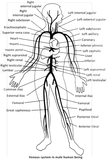
Renal veins : Two, right and left collects blood from respective kidneys and opens into inferior vena cava. The left renal vein is about three times longer than the right one.
Suprarenal vein : Two, right and left, collects blood from adrenal glands. Right one opens into inferior vena cava whereas left one opens into left renal vein.
Inferior phrenic veins : These veins drain the blood from lower surface of diaphragm. The right one ends in post caval. The left one is often doubled with its one branch ending in left renal or suprarenal vein and the other in post caval.
Hepatic veins : They drain blood from liver into the post caval. Urea is maximum in hepatic vein while it is minimum in renal vein.


You need to login to perform this action.
You will be redirected in
3 sec

