Physiology Of Digestion
Category : 11th Class
The process of digestion involves following steps –
(1) Ingestion : It is the intake of food most of the animals capture the prey/food with the help of mouth or tongue.
(2) Mastication : The process occurs in the buccopharyngeal cavity of mammals with the help of teeth. During this process food is broken down into small pieces, which increases its surface area. In frog teeth are not meant for mastication but prevents the escape of prey from mouth.
(3) Deglutition / swallowing : The passage of food from buccopharyngeal cavity to oesophagus/stomach. In mammals bolus of the masticated food is formed in buccopharyngeal cavity which easily slides into oesophagus. It is a voluntary reflex mechanism. Peristalsis is alternative contraction and relaxation of circular and longitudinal muscles produces the wave of contraction due to which the food passes from front to backward direction in the lumen of alimentary canal. The phenomenon is called as peristalsis. Beside alimentary canal, it is also found in vas deference, ureter etc. Peristalsis is maximum in oesophagus and minimum in rectum.
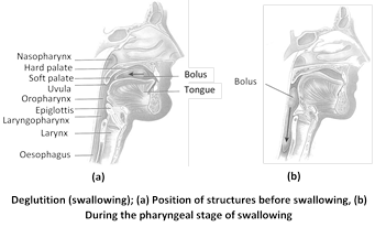
Antiperistalsis is the peristaltic wave occurs in the reverse direction. It occurs in alimentary canal and results in vomiting. The phenomenon is called as “Regurgitation”.
(4) Digestion : The process by which complex food is converted into simple food with the help of digestive enzymes. The process of digestion in mammals starts in buccopharyngeal cavity.
(i) Digestion in buccopharyngeal cavity : In buccopharyngeal cavity of mammals only starch is digested which is 5% of total food or \[20-30%\] of carbohydrates.
(ii) Digestion in stomach : Chiefly proteins is digested in stomach.
(iii) Digestion in small intestine : All three component carbohydrates, proteins and fats digested in small intestine with the help of enzymes secreted by pancreas and intestinal glands.
(5) Absorption : Ingestion and digestion are the first two phases of the physiological processes occuring in the alimentary tract. The third phase is that of absorption by which the digested nutrients are absorb through the wall of gut into blood.
(i) Absorption from the mouth : Normally, there is no absorption from the mouth, but a few drugs may be absorbed into the blood through the mucous membrane, if allowed to dissolve under the tongue, e.g., isoprenaline, glyceryl trinitrate.
(ii) Absorption from the stomach : In the stomach, absorption takes place to a limited degree. The only substances normally absorbed from the stomach are some water, glucose and considerable amounts of alcohol. These substances are absorbed through the walls of the stomach into the venous circulation. Although iron absorption takes place in the small intestine, it is dissolved out of foods most effectively in the stomach in the presence of \[HCl.\]
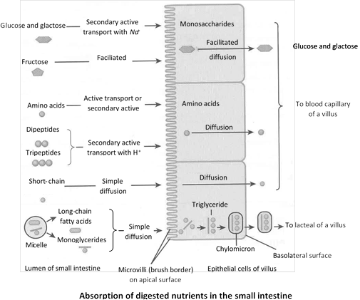
(iii) Absorption from the small intestine : The small intestine is the main absorptive organ. About 90% of the ingested foodstuffs is absorbed in the course of passage through the small intestine.
There are two general pathways for the transport of materials absorbed by the intestine; the veins of the hepatic portal system which lead directly to the liver; and the lymphatic vessels of the intestinal area, which eventually lead to the blood by way of the lymphatic system and the thoracic duct.
Absorption of carbohydrates : The products of carbohydrate digestion is absorbed from the intestine into blood of the portal venous system in the form of monosaccharides, chiefly the hexoses (glucose, fructose, mannose and galactose).
Absorption of amino acids and protein : It is probable that under normal circumstances the dietary proteins are almost completely digested to their constituent amino acids and that these end products of protein digestion are then actively transported from the intestine into the portal blood. Surplus amino acids are also withdrawn from portal blood by liver cells and deaminated into ammonia and keto acids. The ammonia is converted to urea and released into blood for excretion by kidneys, while the keto acids are converted to glucose or pyruvic acid and utilized for energy-production or for storage as glycogen and fat.
Absorption of fats : The dietary fat is digested, by the action of the pancreatic lipase present in the intestine, partially into glycerol and fatty acids and partially to split products such as monoacyl glycerols. These products of fat digestion enter the mucosal cells of the small intestine in the forms of micelles, fatty acids and glycerol.
By the lacteals, the fat is carried to the cisterna chyli (meaning 'the receiver of the chyle') and then by the thoracic (lymph) duct to the left branchiocephalic vein, where it enters the blood. The lymph reaching the thoracic duct from the intestines contains an excess of fat giving it a milky appearance. It is called chyle. In this way, fatty acids and glycerol are eventually brought into the blood stream and so, by a circuitous route, to the liver. In the liver, they are reorganized and recombined to form human fat.
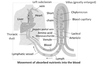
Absorption of vitamins : Water-soluble vitamins like members of B complex (except \[{{B}_{12}}\]) and vitamin C readily diffuse across the walls of the intestine into the blood. The fat-soluble vitamins A, D, E and K are dissolved in micelles, which enter the mucosal cells of the intestine, by simple diffusion. The absorption of these fat-soluble vitamins is markedly decreased in the absence of bile.
(iv) Absorption in large intestine : About 100-200 ml. of the water of undigested food is absorbed in the colon. It helps in maintaining the body water level. Some amount of mineral salts and vitamins are also absorbed. The symbiotic bacteria (E. coli) present in the large intestine, converts the inactive vitamins into active forms (i.e., they synthesizes vitamins (vitamin B complex and vitamin K) which are absorbed.
(6) Assimilation : Conversion of absorbed food into active cytoplasm within cell is called as assimilation.
(7) Faeces formation : The phenomenon occurs in colon due to absorption of water, salts, minerals and vitamins. The peristalsis in colon also helps in faeces formation.
(8) Egestion / defaecation : The elimination of faeces from the elementary canal is called egestion or defaecation. The faeces is waste matter discharged from the alimentary canal.
Pseudo-rumination or coprophagy : Animals swallows night faeces and recycle it through the gut to complete the digestion of cellulose and, making full use of their food. This habit is called coprophagy. Example – Rabbit.
Summary of physiology of digestion Major gastrointestinal enzyme in mammals
|
Name of gland |
Name of digestive juice & optimum pH |
Name of enzyme |
Site of action |
Substrates |
Products |
|
Salivary glands |
Saliva (6.3 - 6.8)
|
Ptyalin / Salivary amylase |
Mouth |
Starch, dextrins, glycogen |
Dextrins, maltose, isomaltose and limit dextrin. |
|
Gastric glands |
Gastric Juice (1-3) |
Pepsin
|
Stomach |
Proteins, casein (Milk) |
Peptones, paracasein (curd). Proteoses |
|
Rennin |
Stomach |
Casein |
Paracasein |
||
|
Gastric lipase |
Stomach |
Fats |
Fatty acid and Glycerol. |
||
|
Liver |
Bile juice (7.6-8.6) |
No enzymes |
Duodenum |
Fat |
Makes the food alkaline, emulsifies fat and kills the harmful bacteria. |
|
Liver |
Bile ( 7.6 - 8.6) |
No enzyme but useful digestive juice, provides alkaline medium, stops the action of HCl. Emulsifies fats and kills harmful bacteria. |
|||
|
Pancreas
|
Pancreatic Juice (8.8) |
Amylase/Diastase |
Small intestine |
Starch, dextrins, glycogen. |
'Limits' dextrins, maltose, isomaltose. |
|
Trypsin
|
Small intestine |
Proteins, Chymotry-psinogen (inactive) procarboxy pept- idases (inactive) Fibrinogen (blood) Casein (milk) |
Peptides, Chymotrypsin (active) carboxy peptidases (active) Elastase (active), Fibrin (clot) Paracasein (curd) |
||
|
Chymotrypsin
|
Small intestine |
Peptones |
Peptides |
||
|
Carboxypeptidases |
Small intestine |
Peptides |
Smaller peptides and Amino acids. |
||
|
Lipase / Steapsin |
Small intestine |
Triglycerides |
Mono-glycerides, fatty acids
|
||
|
DNAase |
Small intestine |
DNA |
Deoxyribonucleotides |
||
|
RNAase |
Small intestine |
RNA |
Ribonucleotides |
||
|
Intestinal glands |
Intestinal Juice (7.5-8.3)
|
Enteropeptidase (enterokinase) |
Small Intestine |
Trypsinogen (inactive) |
Trypsin (active) |
|
Aminopeptidase |
Small Intestine |
Peptides |
Smaller peptides and amino acid |
||
|
Dipeptidases |
Small Intestine |
Dipeptides 'Limit dextrins' |
Amino acids |
||
|
Isomaltase |
Small Intestine |
Isomaltose |
Glucose |
||
|
Maltase |
Small Intestine |
Maltose |
Glucose |
||
|
Sucrase/Invertase |
Small Intestine |
Sucrose |
Glucose, fructose |
||
|
Lactase |
Small Intestine |
Lactose |
Glucose, galactose |
||
|
Lipase |
Small Intestine |
Triglycerides |
Monoglycerides, fatty acids |
||
|
Nucleotidase |
Small Intestine |
Nucleotides |
Nucleosides, inorganic phosphate |
||
|
Nucleosidase Phosphorylases |
Small Intestine |
Nucleosides Phosphate |
Purine, pyrimidine, pentose, phosphate |
||
(9) Hormonal control of digestion : Activities of digestive tract are coordinated by nervous and endocrine systems. Sight and smell of food stimulates nervous system which induces the salivary glands to produce large quantity of saliva, stomach to release its hormone gastrin and intestine to produce intestinal hormones. Other hormones are produced in sequential order. All of them are polypeptide hormones.
Gastrointestinal hormones in mammals
|
Hormone |
Source |
Stimulus for secretion |
Target organ |
Action |
|
Gastrin |
Mucosa of pyloric stomach |
Distension of stomach on food entry |
Stomach |
Stimulates secretion of gastric juice. Constricts cardiac sphincter. |
|
Enterogastrone
|
Duodenal epithelium |
Chyme entry into duodenum |
Stomach |
Slows gastric contractions to delay its emptying. Stops secretion of gastric juice. |
|
Secretin
|
Duodenal epithelium
|
Acidic chyme entry into duodenum
|
Pancreas
Liver Stomach |
Release of sodium bicarbonate in pancreatic juice. Steps up secretion of bile. Inhibits secretion of gastrin. |
|
Cholecystokinin (Pancreozymin) |
Duodenal epithelium |
Presence of fats in duodenum |
Pancreas Gall Bladder |
Release of enzymes in pancreatic juice. Release of bile from gall bladder. |
|
Villikinin |
Intestinal epithelium |
Food in small intestine |
Intestine |
Accelerates movements of villi. |
|
Duocrinin |
Intestinal epithelium (Duodenal mucosa) |
Acidic chyme in intestine |
Intestine (Brunner's gland) |
Release of viscous mucous from Brunner's glands.
|
|
Enterocrinin
|
Intestinal epithelium (Duodenal mucosa) |
Acidic chyme in intestine |
Intestine (crypts of Lieberkuhn's) |
Release of enzymes from Lieberkuhn?s crypts. |


You need to login to perform this action.
You will be redirected in
3 sec

