Eye
Category : 11th Class
Human have binocular vision. The eye can discriminate colour, appraise length, width and depth visually and form true inverted image.
Structure of eye
The eyes are two in number and lodged in orbits (bony socket) of skull. The eye is a hollow, spherical organ, about 2.5 cm in diameter and about 6 to 8 gram in weight. It has two parts -
(1) Protective devices : Eye has four protective devices.
(i) Eye brows : The outwardly directed hair of the eyebrows carry the sweat and rain drops trickling down the forehead to the sides to prevent their falling into the eyes.
(ii) Eye lids (Palpebrae) : In man two eyelids are present, upper is movable. They are regularly closed at short intervals to clean the cornea. This is called blinking. In frog out of two upper eyelid is immovable and lower eyelid is movable. Nictitating membrane is present in frog which protect eye in water. Movement of nictitating membrane takes place by retractor bulbi. It becomes folded by levator bulbi.
A nonfunctional vestigeal nictitating membrane, called plica semilunaris, occurs in human eyes. It remains permanently retracted at the inner angle of each eye.
(iii) Eye lashes : The eyelids bear at the free edge a row of stiff hair, the eye lashes. These check the entry of dust particles, tiny insects and rain drops into the eyes.
(iv) Eye glands
(a) Meibomian gland : The eye-lids bear at the free edge a row of meibomian gland that is modified sebaceous gland. (Act as a lumbricant).
(b) Lacrimal gland or Tear gland : It lies in the upper outer part of the orbit and secretes a slightly saline, watery fluid that contains a bacteriolytic enzyme named lysozyme. This secretion moistens the surface of the eyeball. The excess of this secretion passes through nasolacrimal duct. It is modified sweat gland.
(c) Harderian gland : Some aquatic mammals (whale) possess harderian gland which lubricate nictitating membrane. It is also found in frog and birds.
(d) Glands of zeis (zis) : These are modified sebaceous gland, found at base of hair follicle of eye lashes, pour lubricating fluid in hair follicle. Infection of these glands is sty.
(e) Glands of Moll : It is modified sweat gland and open into the follicles of eyelashes.
In human meibomian, lachrymal, Moll's glands, and zeis glands are present.
(v) Connective tissue : A layer of fatty connective tissue surrounds the eyeball. It serves as a soft shockproof pad.
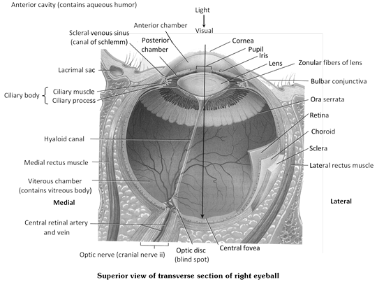
(2) Eye ball : Eye ball is made up of 3 coats or tunic.
(i) Sclerotic layer (Fibrous tunica) : Outer most and opaque, fibrous and non-vascular layer easily seen as white of the eye. It is a coat of dense connective tissue made up of collagen fibers and fibroblasts. Sclera covers entire eye ball except cornea, gives shape to eye ball. Sclera in frog is cartilaginous.
(a) Cornea : In the centre, sclerotic layer it merges with the transparent round window called cornea.
(b) Conjunctiva : The cornea and exposed part of sclera are covered externally by a thin, transparent membrane the conjunctiva.
(ii) Choroid layer (Vascular tunica) : Also known as uvea middle. It is vascular layer which supplies nutrients to the eye. It is distinguished into three parts choroid, ciliary body and iris.
(b) Choroid : It is highly vascular posterior portion of vascular tunic. The choroid occurs in the main part of eye ball adhered to the sclerotic. (The pigment is reddish in rabbit and black, brown or bluish in man).
(b) Ciliary body : Ciliary body is vascular and pigmented like choroid, made up of ciliary processes and ciliary muscles (only circular type). The ciliary body is hidden by iris. The ciliary body helps in accommodation by altering the focus of eye from object or the shape of lens near or far vision.
(c) Iris : Beyond the ciliary body, the vascular tunic sharply turns inwards, forming a circular, shelf-like diaphragm called iris. The colour of the iris is responsible for colour of eye e.g., brown, black, blue or green. In albinos, iris is deficient of pigments.
Lens : Lens is colourless, transparent and fibrous crystaline structure made up of protein (a and b crystalline protein) and enclosed in lens membrane. It is ectodermal in origin. Lens is lodged in eye ball by suspansory ligament of ciliary body. Suspansory ligaments are known as "Zonula of Zinn". In man lens is biconvex while in frog it is elliptical (subspherical).
Lens divide the eye ball in 2 chamber outer aqueous chamber (partially divided into a large anterior and a smaller posterior chamber) filled with aqueous humor (watery) formed by ciliary body and inner vitreous chamber filled with vitreous jelly (or Wharton's jelly) containing 99% water, some salt a little mucoprotein (vitrein) and hyaluronic acid.
Differences between Aqueous humour and Vitreous humour
|
S.No. |
Aqueous humor |
Vitreous humor |
|
1. |
It occurs in aqueous chambers. |
It occurs in vitreous chamber. |
|
2. |
It is a watery fluid |
It is a jelly-like substance. |
|
3. |
It is secreted by ciliary processes. |
It is apparently secreted by retina during development of eye. |
|
4. |
It is continuously absorbed into blood and replaced. |
It is not absorbed or replaced |
|
5. |
It contains most of the diffusible substances of the plasma |
It consists of water (99%) protein vitrein, hyaluronic acid and collagen fibres. |
|
6. |
Obstruction in its flow may damage retina by increasing intraocular pressure. |
It does not flow. |
|
7. |
Refrective index is 1.33 D |
1.34 D |
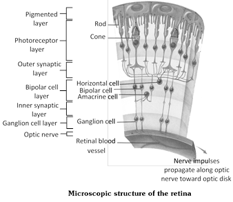
(iii) Retina (Neurosensory tunica) : It is innermost, thin and transparent, purplish red due to the present of the eye pigment rhodopsin (in rods) or visual purple which was extracted by Kuhne (1876) and named 'Schpurpur' (Visual purple). Made up of 4 distinct layer -
(a) Cuboidal pigmented epithelium (towards choroid).
(b) Layer of rods and cones.
(c) Layer of bipolar neurons.
(d) Layer of ganglia (Towards vitreous chamber innermost).
Area centralis of retina : A little part of retina that lies upon the optical axis is called area centralis. Here, the retina is very thin and contains only cone cells filled with a yellow pigment. Hence, this part is called yellow spot or maculla lutea. In man (Rabbits) and other mammals, but not in frogs, this area has a small shallow dispression called fovea centralis. The latter is the most sensitive part of an eye, i.e. the area of most acute vision. It is also claimed that the cone cells in fovea centralis are placed somewhat obliquely. So that these can form magnified images of object.
Blind spot (Optic disc) : At this point, the optic nerve turns towards the outer side, pierces through the whole thickness of the wall of eyeball, forming an optic foramen and runs to the brain. Obviously, the region of optic foramen has no retina. It therefore, does not take part in image formation and is called blind spot.
Differences between Blind spot and Yellow spot
|
Blind spot (Optic disc) |
Yellow spot (Macula lutea) |
|
It lies a little away from the yellow spot. |
It lies exactly opposite the centre of the cornea. |
|
It contains no pigment. |
It has a yellow pigment. |
|
Optic nerve starts from this spot. |
No nerve starts from this spot. |
|
It lacks a depression. |
It has a shallow depression, the fovea centrallis, at its middle. |
|
It lacks visual receptors and is insensitive to light. |
It has visual receptors and is sensitive to light. |
|
The eye coats are absent at blind spot. |
Eye coats are present at yellow spot. |
|
No image is formed at this place. |
Image is formed at this place. |
Ora seratta : The function retina terminates anteriorly along an irregular border, the ora seratta.
Function of the parts of human eye
|
Part |
Function |
Part |
Function |
|
Lens |
Refracts and focuses light. |
Ciliary body |
Holds lens in place. |
|
Iris |
Regulates light entrance. |
Retina |
Contains receptors. |
|
Pupil |
Admits light. |
Rods |
Allow black and white vision |
|
Choroid |
Absorbs extra light. |
Cones |
Allow colour vision. |
|
Sclera |
Protects |
Optic nerve |
Transmits impulse. |
|
Cornea |
Refracts light. |
Fovea centralis |
Region of cones in retina |
|
Humors |
Refracts light. |
|
|
Working of eye
(i) Mechanism of light perception : The human eye has two functional parts – Dioptric or Focussing part and Receptor part.
(ii) Focussing part : It consists of conjunctiva, cornea, aqueous humour lens and vitreous humour. These part are transparent and act as lenses. They refract the light rays passing through the eye to bring them to a focus on the retina. Maximum refraction is caused by the cornea, which places the image approximately on the retina. The lens effects fine adjustment and brings the image into a sharp focus.
(iii) Receptor part : It comprises the retina. The image formed on the retina is inverted and smaller. It converts the energy of specific wave lengths of light into action potential in nerve fibre.
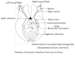
(a) Pathway of sensory impulses from eye to brain : The nerve impulses generated in the retina of the eye in response to light follow a definitive path and terminates in visual cortex in each optic lobe which act as primary visual center.
Biochemistry of eye
The receptor cells of eye are called photoreceptor or visual cells. They are of two types - Rod cells and Cone
(1) Rod cells : The rod cells contain a purplish pigment called visual purple or rhodopsin. They function in dim light and at night. They produce poorly defined images. Bright light splits rhodopsin into a lipoprotein scotopsin and a carotenoid pigment retinal (retinine) a process called bleaching. The spiliting of rhodopsin depolarizes the rod cell and it releases a neurotransmitter, passing the nerve impulse via bipolar neuron and ganglion cells to the optic nerve. In the dark, rhodopsin is resynthesized from scotopsin and retinal. This process is called 'dark adaptation'. It makes the rods functional. It takes sometime for rhodopsin to be reformed. This is why on entering a dark room at daytime or on coming out of a well lighted room at night we feel blind for a while, when we go from darkness into bright light, we feel difficulty in seeing properly for a moment till rhodopsin is bleached and cones become functional.
(2) Cone cells : Cones contain iodopsin which is visual violet and made up of photopsin + retinal. The 3 types of cones are erythrolobe (775 nm sensitive tored), cyanolabe (430 nm sensitive to blue) and chlorolabe (sensitive to green 535 nm). However, if all the cone, types are simultaneously stimulated by equal amounts of coloured light than sensation for white light is perceived.
Diurnal animals are adapted to see during day light (Photopic vision) and can perceive colour. In dark, colours are not perceived. Such animals have more cones in their eye than rods.
Differences between Rod cells and Cone cells
|
Rod cells |
Cone cells |
|
Rods secreted by rod cells. |
Cones secreted by cone cells |
|
Produce "Rhodopsin" which is visual purple and made up of scotopsin+11 cis retinal. Vitamin-A is needed for the formation of Rhodopsin. |
Produce "Idopsin" which is visual violet and made up of photopsin+11 cis retinal. |
|
Rhodopsin is very sensitive to light. |
It is sensitive to colour. |
|
Rods are active in dim light or low intensity light. |
Cones are active in bright light which is called photopic vision. |
|
Rod cells are absent in fovea centralis of retina. |
In fovea centralis only cone cells are present. |
|
Rods are more in number in peripheral region of retina. |
Cones are more in number in central region of retina. |
|
Rods are more in nocturnal animals. |
Cones are more in diurenal animals. |
|
In owl only rods are present and cones are absent. |
In fowls only cones are present and rods are absent. |
|
120 million rodsin in human. |
6 million cones in human. |
Accommodation and types of vision
(1) Accommodation : Light passes through many refractive surfaces before it is focussed on the retina forming an inverted and true image. The main sites of refraction are cornea\[\to \]aqueous humor – iris – lens (position can be altered by ciliary body : accommodation) - posterior chamber (= vitreous humor)\[\to \]retina ( in fovea). The refractive index of the eye varies from 59 diopter (when the lens is at rest) to about 71 diopter (when lens is bulging in maximum accomodation). The accommodation reflex occurs when the eye changes its focus from a far away object to nearer one. The change in strength of the lens provides the physiological basis of accommodation. Radial and circular muscle fibres of ciliary muscles play an important role in this as they contract reflexively (parasympathetic control) and increase lens strength. The pupil constricts. This facilitates increase in sharpness of image. Ageing causes loss of accomodation.
Relationship between structures during accommodation
|
Object |
Ciliary muscle |
Suspensory ligament |
Lens |
Refraction |
|
Near |
Contracted |
No tension (Relaxed) |
Thick |
Increased |
|
Distant |
Relaxed |
Tension maximum |
Thin |
Decreased |
(2) Types of vision
(i) Binocular vision : Man has binocular vision in which both the eyes are focussed on the same object but from slightly different angles. The visual fields of both eyes overlap and the foveae of both are focused on the same object. This provides depth to the images, i.e., gives stereoscopic or 3D effect and enables man to judge distances correctly.
(ii) Vision in other animals : Primates and predatory animals, such as owl and cat, have binocular vision. In some animals, such as rabbit, birds, each eye is focussed on a separate object. This is termed monocular vision.
(iii) Colour vision : It is the ability of some animals to detect colours in an object. Humans, apes, monkeys, and most fishes, amphibians, reptiles and birds have strong colour vision. The insects and crayfish also have colour vision. In vertebrates, colour vision results from the activity of cone cells. Most domestic and nocturnal mammals and sharks lack colour vision. They probably see objects in shades of grey (monochrome vision).
(iv) Nocturnal and Diurnal vision : Man has both day vision and night vision as he has both rods and cones in considerable numbers in the retina. Most birds have only day vision as their retina contains mainly cones. Owls have much better night vision than day vision for they possess a large number of rods and few cones in their retina.
Range of vision : The visible range of spectrum varies in animals. Bees, ants, spiders and goldfish can see ultraviolet light, which is invisible to man.
Correspondence between Camera and Eye
|
S.No. |
Camera |
Eye |
|
1. |
Box |
Sclera |
|
2. |
Black inner paint |
Choroid |
|
3. |
Shutter |
Eyelids |
|
4. |
Diaphragm |
Iris |
|
5. |
Light hole |
Pupil |
|
6. |
Lens |
Lens |
|
7. |
Light-sensitive plate or film |
Retina |
|
8. |
Image small and inverted |
Image small and inverted |
Eye movement
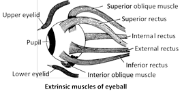
In eye orbit eyeball remain attached with 6 extrinsic muscles.
Out of six, first four are rectus and last two are oblique muscles.
(1) Anterior rectus or Internal ractus
(2) Posterior rectus or External ractus
(3) Inferior rectus
(4) Superior rectus
(5) Inferior oblique muscle
(6) Superior oblique muscle
Eye defects
(1) Myopia
(i) Also known as near sightness.
(ii) Short sightness.
(iii) Near object is clear. Far object is not clear.
(iv) Eyeball become longer.
(v) Image is formed before retina. Can be removed by concave lens.
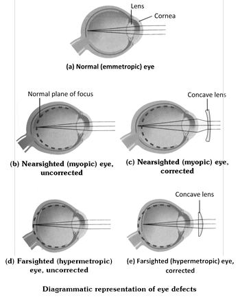
(2) Hypermetropia
(i) Also known as hypermetropia or long sightness.
(ii) Far sightness.
(iii) Far object is clear, near object is not clear.
(iv) Eye ball become short.
(v) Image is formed behind the retina.
(vi) Can be removed by convex lens or lens convient.
(3) Astigmatism
(i) Curvature of cornea become irregular and image is not clearly form.
(ii) Can be removed by cylindrical lens.
(4) Cataract
(i) It is due to defective protein metabolism.
(ii) During this lens or cornea sometime both become opaque.
(iii) Operation is needed.
(5) Gloucoma
(i) It is due to increase in intraocular pressure in aqueous chamber.
(ii) Operation is needed at early stage due to blockage of schlemm’s canal.
(6) Trachoma
(i) It is increased in redness of eye and more secretion of watery fluid.
(ii) It is due to infection of bacteria, chlamidia trachamastis.
(iii) Due to this follicles may form in conjunctiva.
(7) Xerothalmia
(i) It is due to deficiency of vitamin A. \[({{A}_{2}})\]
(ii) During this conjunctiva or cornea becomes keratinized.
(iii) It may lead to blindness.
(8) Strabimus
(i) In this type eyeball remain in some what in bended position.
(ii) It is due to long extra ocular muscles during development of eye.
(iii) Operation is needed at early stage.
(iv) Also associated with squint.
(9) Presbiopia
(i) During this power of accommodation of lens decreases due to age factor and defected metabolism.
(ii) Also known as age sightness.
(iii) Can be removed by bifocal lens.
(10) Photofobia : No clear image in bright light.
(11) Emmetropia : Normal vision.


You need to login to perform this action.
You will be redirected in
3 sec

