Phonoreceptor And Mechanism Of Hearing Or Auditory Sensations And Equilibrium
Category : 11th Class
Also known as stato-acuostic organ. It is the receptor for balancing and hearing which is sensitive for gravity and sound waves. It is also sensitive in orientation of body. It is also known as mechano receptor because of it change mechanical energy of sound waves in to action potential.
Structure of Ear
Ear of mammal is divided in to 3 parts -
(1) External ear : It is made up of pinna and auditory meatus. Pinna is found in only mammals. Its upper rounded part is helix and lower is ear lobe. It is made up of adipose connective tissue and elastic cartilage and has ear muscles which are vestigeal in case of human beings. Pinna collect the sound waves and drive towards auditory meatus.
Auditory meatus is 25 mm. long canal lined by simple columnar epithelia and made up of fibro elastic cartilage. It possesses ceruminus gland which secrete cerumin (ear wax). Cerumin trap the dust particles and microbes.
Tympanic membrane : It is also called ear drum and present at the junction of auditory meatus and tympanic cavity.
(2) Middle ear : The cavity of middle ear is known as tympanic cavity which is enclosed by tympanic bulla bone of skull and filled with air. Middle ear separated from external ear by ear drum and from internal ear by thin bony portion or partition with two openings known as oval and round window.
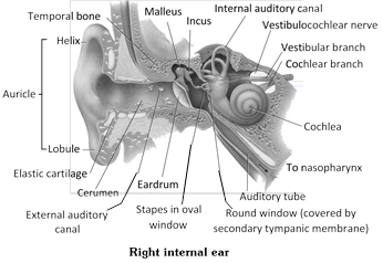
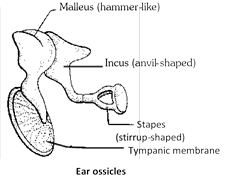
(i) Ear ossicle : A chain of three small, movable bones, the auditory or ear ossicles crosses the tympanic cavity. The outer ossicle is attached to the inner surface of the tympanic membrane.
Ear ossicles
|
Ear ossicle |
Shape |
Modification of |
|
M - Malleus |
Hammer shaped |
Articular bone of lower jaw. |
|
I - Incus |
Anvil shaped |
Quadrate bone |
|
S - Stapes |
Steirrup shaped |
Hyomandibular of columella |
In man ear ossicles are known as H.A.S. stapes is the smallest bone of the body. In frog only stapes is present.
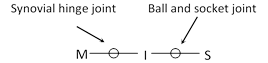
(ii) Joints
(iii) Muscles
Tencer tympani : Limits movements and increases tension on eardrum to prevent damage to inner ear from loud sound.
(iv) Eustachian tube : It is made up of elastic cartilage and it connect middle ear to nosopharynx. It maintain equilibrium in and out side of the tympanic membrane. Blocking of eustachian tube impairs hearing due to imperfect vibrations of drum. Eustachian tube is normally closed, it opens during swallowing and yawning.
(v) Fenestrae : Between middle ear and internal ear a thin bony membrane is present which possess two apertures (Windows).
(a) Fenestra ovalis : It is upper window, connect middle ear to internal ear and guarded by membrane. End of stapes is fit on the upper window. It is towards vestibule so it is also known as F. vestibuli.
(b) Fenestra rotundus : It is ventral window, connect middle ear to internal ear and guarded by membrane. It is towards scala tympani so it is also known as F. Tympani (also known as F. cochleae).
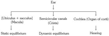
(3) Internal ear (Membranous labyrinth) : Internal ear is also known as membranous labyrinth and enclosed by bony labyrinth. Bony labyrinth is formed by periotic bone or petrous. A cavity is present between membranous labyrinth and bony labyrinth known as perilymphatic space. It is filled with perilymph and endolymph is found in membranous labyrinth. The membranus labyrinth consists of 2 parts - Vestibule and Cochlea.

(i) Vestibule : The vestibule is a central sac like part. It further consists of 2 chambers large - Utriculus (Upper) and smaller - sacculus (lower).
(a) Semicircular canal : From utriculus 3 semicircular canals arise these are –
Anterior semicircular canal (Superior)
Posterior semicircular canal (Inferior)
Horizontal semicircular canal (External)
They are perpendicular each other.
Crus commune : A common part of anterior and posterior semicircular canal arise from dorsal region of utriculus is known as crus commune.
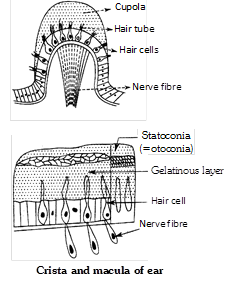
Ampulla : Terminal part of the each semicircular canal is enlarged to form an ampulla.
Crista : Each ampulla has a sensory spot called crista ampullaris or simply crista, for equilibrium.
(b) Sacculus : It is a lower chamber of vestibule. From the lower part of the sacculus arises a short tube, the ductus reuniens, that joins the cochlear duct.
Ductus endolymphaticus : It is filled with endolymph and arises from the junction of utriculus and sacculus.
Macula : are present in utriculus and sacculus. it is a group of sensory cells. In man (Rabbit) 2 maculas are present. (A crista resembles a macula in structure except that lies on an elevation, the acoustic ridge, its sensory cells have longer "hair", and its gelatinous mass is dome shaped, lacks otoliths and is called cupula.)
Difference between Crista and Macula
|
S.No. |
Crista |
Macula |
|
1. |
Found in ampulla of semi-circular canal |
Found in vestibule i.e. sacculus and utriculus. |
|
2. |
Their total number is 3 |
Only 2 are present |
|
3. |
No otolith |
Otolith present |
|
4. |
Long auditory hairs |
Short auditory hair |
|
5. |
Facilitate maintenance of dynamic equilibrium and angular acceleration e.g. rotational movement of head |
Help in static equilibrium and linear acceleration e.g. tilting of head or body at rest and rapid forward movement. |
Otolith : Also known as otoconia made up of protein and calcium carbonate and present in endolymph.
(ii) Cochlear duct and Cochlea : It is a spirally coiled tube (\[23\]coiling) which is connected to sacculus by a short duct. It is divided into 3 chambers by 2 membranes.
(a) Scala vestibuli : Upper chamber filled with - perilymph - connect with middle ear by F. ovalis, or oral window.
(b) Scala media (Real cochlear duct) : Middle chamber filled with - endolymph.
(c) Scala tympani : Lower chamber filled with - perilymph connect with middle ear by F. Tympani or round window.
(d) Reissner's membrane : Present at the roof of scala media, it saparate S.M. to S.V.
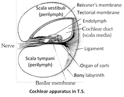
(e) Basilear membrane : Present at the base of S.M. It is thicker than Reissner's membrane and it separates S.M. to S.T.
(f) Modiolus : A bony core around which bony spiral canal of cochlea make \[2\frac{3}{4}\] turns or coils in man.
(g) Helicotrema : A aperture present in scala media which connect scala vestibuli to scala tympani is known as helicotrema.
(h) Tectorial membrane : Tectorial membrane is a leaf like gelatinous structure present at the dorsal side of organ of corti.
(i) Organ of corti : Discovered by Italian anatomist Alfanso-Corti. Also known as ridges of corti which are present in basiler membrane. Organ of Corti contains a variety of cells. They receives nutrients from endolymph. The cells of organ of Corti are following types -
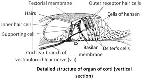
Characteristics of cells of organs of Corti
|
Receptor cell (=Hair cells) |
Supporting cells |
Tectorial membrane |
Peripheral membrane |
|
Two type (i)- inner hair cells - in one layer and number 3500, while the (ii) outer hair cells are in 3 - 4 rows (20,000) |
Support hair cells, These rest on basilar membrane |
Flap of fibrous and gelatinous tissue, the outer right plate is called reticular lamina which is supported by rods or corti anchored to basilar membrane |
Has restricted elasticity respond to low to high frequencies within audible region |
|
Hair of outer hair cell extend into scala media and embeded in roof like tectorial membrane. |
Provide nutrients and physical support to the hair cells |
|
|
|
Inner hair cells respond to the velocity of movement of the basilar membrane. While the outer hair cells are primarily concerned with the displacement of the basilar membrane by the sound waves. |
They are nut involved in sound transduction |
|
|
|
Hair cells have a basal body just under the hair. The basal body facilitates transduction of the mechanical signal to a neural signal (electrogenesis) |
|
|
|
Mechanism of sound perception
Vone Beskey won the Nobel prize for his work on ear. The mechanism found in ear involve two unrelated functions : Hearings and equilibrium.
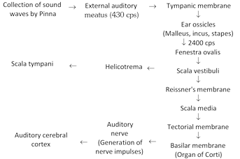
(1) Hearing : The ear not only detects sound but also notes its direction, judges its loudness and determines its pitch (frequency) sound waves are collect by the pinna and directed inward through the external auditory meatus (frequency 430 cycle per second). Here they strike the tympanic membrane. The latter begins to vibrate at the same frequency as that of the sound waves. From the tympanic membrane, the vibration are transmitted across the tympanic cavity by the ear ossicles to the membrane of the fenestra ovalis. The force of vibrations is considerably increased in the middle ear by leverage of the ossicles and also by much smaller surface area of the membrane of fenestra ovalis than that of the tympanic membrane. (The frequency is 2400 cycle/sec). Increase in frequency is important because the sound wave are transmitted from air to a fluid medium. The membrane of fenestra ovalis transmits the vibrations to the perilymph of the scala vestibuli and hence via Reissner's membrane to the endolymph in the scala media. From here the vibrations are transferred to the basilar membrane and the perilymph in the scala tympani.
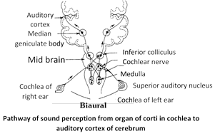
Vibration of the endo lymph of the scala media cause the basilar membrane of this chamber vibrate. Vibrations of the basilar membrane make the "sensory hair" of receptor cells in the organ of corti move in the overlying gelatinous membrane (Tectorial membrane) and get distorted. This stimulation causes depolarisation of the receptor cells and initiation of nerve impulse in the fibres of the auditory nerve. The nerve impulse travels via relay centers e.g. spiral ganglion\[\to \]cochlear nuclei\[\to \]superior auditory nuclei\[\to \]inferior colliculi\[\to \]auditory cortex of cerebrum (The cerebral cortex interprets the impulses as sound). The various steps in the mechanism of hearing
Human ear can hear a frequency of 500 to 5000 hertz (Hz; 1 Hz = 1 cycle/second). However, it can hear the complete range of frequencies from 20 – 20,000 Hz only with intense sound. Sound energy is measured in terms of units called decibels (dB). Sounds in our city homes average \[4050\,\,dB,\] but street noise averages \[7080\,\,dB.\] Sounds up to 80 dB are considered bearable by man, but higher sound intensity are hazardous, causing nervous stress, irritability, increased blood pressure etc. Non stop noise of 90 or more dB produces temporary deafness. 160 dB sound can cause total deafness by rupturing our ear drum. Sound becomes uncomfortable to normal ear at about 120 dB.
(2) Equilibrium : Sound become painful above 140 dB. Exposure to certain antibiotics, such as gentamycin some anticancer drugs, loud sound, loud music, or engine rear of jet planes, vacuum cleaners, damages hair cells of cochlea.
(i) Static equilibrium and linear acceleration : Maculae detect changes in the head (or body) with respect to gravity (static equilibrium) and in the movement in one direction (linear acceleration). With a change in the position of the body, the otoliths, being heavier than the endolymph, press upon the sensory hairs of the maculae. This stimulates the sensory cells which initiates nerve impulse in the fibres of the auditory nerve. The macula of utricle responds to vertical movements of the head, and the macula of saccule responds to lateral (sideways) movement of the head.
On rapid forward movement, the otoliths, because of having greater inertia than the surrounding endolymph, lag behind and press back the sensory hair, stimulating the sensory cells to generate nerve impulses.
(ii) Dynamic equilibrium : Cristae detect turning or rotational movements of the head (angular acceleration). When the head is turned, the endolymph in the semicircular ducts, due to its inertia, does not move as fast as the head and the sensory cells of the crista, but continues to move after the head stops moving. Because of this difference in the rate of movement, the sensory hair of the cristae are swept through the endolymph and become bent over. This disturbance stimulates the sensory cells and sets up action potential in the fibres of the auditory nerve, which transmits it to the brain. Since the three semicircular ducts are arranged in three different planes, a movement of the head in any direction will stimulate the sensory cells of at least one crista.
Defects of ear
(1) Labyrinthine disease : Malfunction of inner ear.
(2) Meniere's disease : Loss of hearing due to defect in cochlea.
(3) Otitis media : Acute infection of middle ear.
(4) Eustachitis : Inflammation of eustachian tube.
(5) Myringitis (Tymanitis) : Inflammation of eardrum.
(6) Otalgia : Earache (pain in ear)


You need to login to perform this action.
You will be redirected in
3 sec

