Cleavage
Category : 12th Class
Definition : The term cleavage refers to a series of rapid mitotic division of the zygote following fertilization, forming a many celled blastula. The cleavage follows fertilization and ends with the formation of a characteristic development stage called blastula.
Cleavage versus typical mitosis : The cleavage division are no doubt mitotic as they produce diploid cells, they differ from typical mitosis in a couple of significant points.
Different between cleavage and mitosis
|
S.No. |
Characters |
Cleavage |
Normal mitosis |
|
1. |
Site of occurrence |
In zygote or parthenogenetic egg |
In most of somatic cells |
|
2. |
Interphase |
Of shorter period |
Of longer period |
|
3. |
Growth |
Does not occur |
Occurs during interphase |
|
4. |
Oxygen consumption |
High as is very rapid process |
Low as is slow process |
|
5. |
Size of daughter cells |
Decreases |
Remains same after growth |
|
6. |
DNA synthesis |
Faster |
Slower |
|
7. |
Nuclear-cytoplasmic ratio |
Increases |
Remain same |
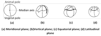
Planes of cleavage : The cleavage is initiated by the appearance of a constriction or groove called cleavage furrow. The cleavage furrows may divided the egg from different angles or planes. These are four important planes of cleavage. They are as follows.
(1) Meridional plane : When cleavage furrow bisects both the poles of the egg, passing through the animal vegetal axis, the plane of cleavage is called meridional plane.
Example : Ist and IInd cleavage furrow of frog and Ist cleavage furrow of chick.
(2) Vertical plane : When cleavage furrow passes from the animal pole to the vegetal pole, but it does not pass through the median axis of the egg.
Example : IIIrd cleavage furrow of chick, Amia calva and Lepidosteus.
(3) Equatorial plane : When cleavage furrow bisect the egg at right angles to the median axis and half way between the animal and vegetal poles.
Example : Ist cleavage plane of eggs of higher mammals and vth cleavage plane of the eggs of Ambystoma maculatum.
(4) Latitudinal or transverse or horizontal plane : The transverse plane resemble the equatorial plane, but it passes either above (towards the animal pole) or below (towards the vegetal pole) the equator of the egg.
Example : IIIrd cleavage plane of Amphioxus and frog.
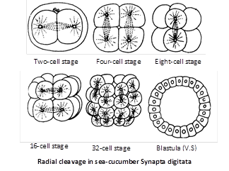
Patterns of cleavage : During segmentation, the cleavage furrows are not formed at random but are oriented in a particular manner with reference to the main (animal-vegetal) axis of the egg. The orientation of successive cleavage furrows with respect to each other and to the main axis of the egg is, however, unlike in different species. As such various patterns of cleavage are found among animals. Based upon symmetry, four patterns of cleavage have been recognized. They are as follows
(1) Radial cleavage : In this cleavage pattern, division take place in such a manner that all the blastomeres are placed in a radially symmetrical fashion around the polar axis. When such an egg is viewed from the poles, the blastomeres seem to be arranged in a radially symmetric form.
Example : Sponges, coelenterates, sea urchin, sea cucumber, amphioxus.
(2) Biradial cleavage : In this pattern four blastomeres arise by the usual two meridional cleavages. The third cleavage plane is vertical resulting in the formation of a curved plate of 8 cells arranged in two rows of 4 each. In these rows, the central cells are larger than the end ones.
Example : Ctenophores like Beroe.

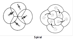
(3) Spiral cleavage : The spiral cleavage is diagonal to the polar axis. In this type, the spindles for the third cleavage, instead of being erect, are oriented diagonally so that the resulting upper tier of cells is sidewise. The upper 4 cells are placed over the junction between the four lower cells. The upper smaller cells are called micro and lower larger cells are known as macromeres. The spiral cleavage results due to oblique positions of the mitotic spindles. This type of cleavage is called the spiral type because the four spindle during the third cleavage are arranged in a sort of spiral.
Examples : Eggs of annelids, molluscs, nemerteans and some of the planarians.
(4) Bilateral cleavage : In this pattern of cleavage, the blastomeres are so arranged that the right and left sides becomes distinct. In this case, two of the first four blastomeres may be larger than the other two, thus establishing a plane of bilateral symmetry in the developing embryo.
Examples : Nematodes, cephalopodes, molluscs, some echinoderms, tunicates, Amphibia and higher mammals.

Laws of cleavage : Certain fundamental rules or laws of cleavage are following :
(1) Sach’s laws : In 1877, Sach proposed following two laws :
(i) Cells tend to divide into equal daughter cells.
(ii) Each new division plane tends to intersect the preceding plane at right angles. (Acts to maintain the spheroidal shape of blastomeres).
(2) Hertwig’s laws : In 1881, O. Hertwig added following laws of cleavage in Sach’s laws :
(i) The nucleus and achromatic figure (or mitotic spindle) ‘occupy the centre of protoplasmic density’ of the egg or blastomeres in which it lies. Hence, in the microlecithal and isolecithal eggs, the spindle is located centrally; in a telolecithal ovum it is nearer the animal pole.
Corollary : Blastomeres divide into two equal parts unless the yolk is unevenly stored in them.
(ii) The axis of a mitotic spindle occupies the longest axis of the protoplasmic mass in which it lies and division therefore tends to cut this axis transversely (evident in ovoid blastomeres).
Corollary : The ensuing plane of division cuts across the long axis, and the daughter cells revert to a more spheroidal shape.
(3) Balfour’s law : Balfour’s law which was formulated by Balfour in 1885, states that the speed or rate of cleavage in any region of egg is inversely proportional to the amount of yolk or deutoplasm it contains. (In telolecithal eggs, blastomeres at the animal pole divide faster than those nearer the vegetal pole).
(4) Pfluger’s law : The mitotic spindle always elongates in the direction of least resistance.
Types of cleavage : The amount of yolk (Lecithality) also determines the type of cleavage. Which are as follows
(i) Holoblastic cleavage : Alecithal, homolecithal and mesolecithal eggs show rapid and complete division of zygote are called total or holoblastic cleavage. Resulting 8 blastomeres after the third cleavage may be equal or unequal to each other. Accordingly they are of two types
(a) Equal holoblastic cleavage : If the blastomeres are approximately equal, it is called equal holoblastic cleavage.
Examples : Echinoderms, amphioxus and placental mammals.
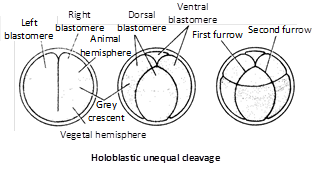
(b) Unequal holoblastic cleavage : If the upper 4 blastomere are smaller (micromeres) than the lower 4 yolk-laden larger blastomere (macromere), it is called unequal holoblastic cleavage.
Example : Fish and amphibians.
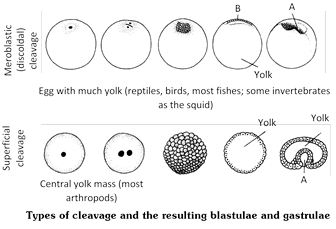
(ii) Meroblastic cleavage : In large polylecithal eggs cleavage furrow cannot cut through the enormous yolk present so that the entire egg is not divided into cells. Thus cleavage is incomplete or partial, termed meroblastic. It is of following two types
(a) Discoidal cleavage : Cleavage are restricted only to the small cytoplasmic cap at the animal pole resulting in a rounded embryonic or germinal disc is termed discoidal cleavage.
Example : Eggs of elasmobranchs, bony fishes, birds, reptiles and egg laying mammals.
(b) Superficial cleavage : Cleavage is restricted to a superficial peripheral layer of cytoplasm around yolk, hence the term superficial cleavage.
Example : Centrolecithal eggs of arthropods.
Cleavage in human zygote
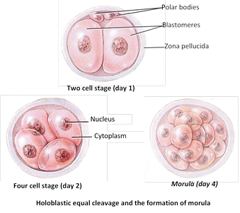
Cleavage in the human zygote occurs during its passage through the fallopian tube to the uterus as in other mammals. It is holoblastic. The first cleavage takes place about 30 hours after fertilization. It is meridional, coinciding with the animal-vegetal pole axis. It produces two blastomeres, one slightly larger than the other. The two blastomeres remain adhered to each other. The second cleavage occurs within 60 hours after fertilization. Third cleavage takes place about 72 hours after fertilization.
(1) Formation of morula : After 4th cleavage solid ball consist of 16 to 32 cells are formed which looks as a little mulberry called morula. Due to holoblastic and unequal cleavage, two types of blastomere are formed.
There is an outer layer of smaller (micromere) transparent cells around on inner mass of larger cells (macromere). The morula reaches the uterus about 4 to 6 days after fertilization. It is still surrounded by the zona pellucida, that prevents its sticking to the uterine wall.
(2) Formation of blastula (blastocyst) : The outer layer of cell becomes that and form trophoblast or trophoectoderm which draws the nutritive material secreted by the uterine endometrial glands. The fluids absorbed by the trophoblast collects in a new central cavity called blastocoel.
As the amount of nutritive fluid increases in blastocoel, morula enlarges and takes the form of a cyst and is now called blastocyst or blastodermic vesicle. The cells of trophoblast do not participate in the formation of embryo proper.
Inner cell mass of macromeres forms a knob at one side of trophoblast and forms an embryonal knob and is primarily determined to form the body of developing embryo so is called precursor of the embryo.
Types of blastula
(i) Coeloblastula : A hollow blastula in which blastocoel is surrounded by either single layered (e.g., echinoderms, amphioxus) or many layered blastoderm (e.g., frog).
(ii) Amphiblastula : It is a type of coeloblastula surrounded by two types of cells. Upper micromeres and lower macromeres. Ex. : frog.
(iii) Stereoblastula : Solid blastula with no blastocoel e.g., in coelentrates annelids and molluscs.
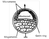
(iv) Discoblastula : The blastula is as a multilayered flat disc at the animal pole lying on the top of well developed yolk. It is found in reptiles, birds, prototherians and fishes.
(v) Blastocyst : In this, the blastula is as a cyst with 2 types of cells : an outer epithelium – like layer of trophoblast or nutritive cells; and an inner mass of formative cells collectively called embryonal knob.
(vi) Superficial blastula or periblastula : In this, the blastocoel is filled with yolk and is surrounded by a peripheral layer of cells. It is found in insects.
Fate mape :
Fate mapping Ist tried by SPECT (1919)
Fate mapping Ist done by W. Vogt. (1929)
Fate mapping is done by vital dyes.
e.g., Neutral Red. Nile blue sulphate, Bismarck brown.
Stain carrier = Agar-Agar and cellophan
Fate mapping can be done in Later blastula or earlygastrula.
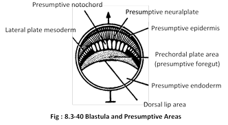
Presumptive Areas : Also known as prospective areas.
(1) Animal pole : Animal pole gives rise presumptive ectoderm. It is the region of presumptive epidermis + presumptive neural plate.
(2) Dorsal surface : On dorsally a small area lies in intermediate zone which is the area of Presumptive Notochord.
(3) Laterally : Closed to the notochord, presumptive mesoderm is present.
(4) Vegetal half gives rise presumptive endoderm.


You need to login to perform this action.
You will be redirected in
3 sec

