Gastrulation
Category : 12th Class
Definition : Gastrulation is a dynamic process involving critical changes in the embryo such as differentiation of cells, establishment of the three primary germ layers and transformation of the single walled blastula into a double walled gastrula.
Types of gastrular movement or morphogenetic movement : The movements of cells during gastrulation is called formative or morphogenetic movements. Following types of gastrular movements are found in different animals
(1) Epiboly : It involves the morphogenetic movement of prospective ectodermal (micromeres) blastomeres antero-posteriorly to envelop the presumptive endodermal and mesodermal blastomeres. It is found in telolecithal egg of frog.
(2) Emboly : It involves inward movement of prospective endodermal and chorda-mesodermal blastomeres from the surface of blastula. Emboly includes following methods :
(i) Invagination : It involves insinking of endodermal cells in the blastocoel to form archenteron. It is found in amphioxus.
(ii) Involution : It involves the rolling in of the chorda-mesodermal blastomeres inside the ectodermal cells over the lips of blastopore. It is also found in the gastrulation of frog.
(iii) Ingression or polyinvagination : In this, individual blastomeres migrate into the blastocoel either from only vegetal pole (called unipolar ingression e.g., Obelia;) or from all sides (called multipolar ingression e.g., Hydra) to form a solid gastrula called stereogastrula.
(iv) Delamination : It involves splitting off the blastoderm into two layers by the appearance of grooves resulting the formation of hypoblast. It is found in birds.
Formation of layers by gastrulation : Gastrulation includes the formation of following structures
(1) Formation of endoderm : The blastodermic vesicle enlarges and cells present on the lower surface of the embryonal knob detach by delamination from the embryonal knob. The part of endoderm located under the embryonal knob is called embryonic endoderm which later forms embryonic gut, while the remaining part of endoderm along with trophoblast forms the yolk sac.
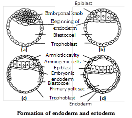
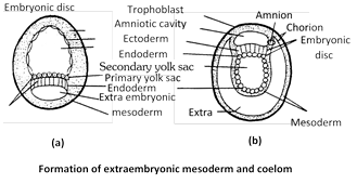
(b) Formation of embryonic disc and mesoderm : Meanwhile, the blastocyst continues to grow due to absorption of more and more uterine milk. The embryonal knob stretches and cells of Rauber start breaking off and dispersing. So the cells of embryonal knob from a regular layer called embryonic disc which becomes continuous with the trophoblast. Embryonic disc is differentiated into cephalic, embryonic and caudal regions. Formation of embryonic mesoderm starts at the caudal region of the embryonic disc where cells undergo rapid proliferation and form a localized thickening of the embryonic disc and form the mesodermal layer between ectoderm and endoderm.
(3) Formation of ectoderm : The remaining cells of blastodisc become columnar and form ectoderm.
Fate of germ layers : Each of the three germ layers gives rise to definite tissues, organs and systems of the body. Their fate in embryo and adult has been listed below.
Fate of germ layer
|
Ectoderm |
Mesoderm |
Endoderm |
|
Epidermis and skin derivatives |
Dermis |
Gut |
|
Cutaneous gland |
Muscular tissue |
Glands of stomach and intestine |
|
Nervous system (Brain + spinal cord) |
Connective tissue |
Tongue |
|
Motor and optic nerve |
Endoskeleton |
Lung, trachea and bronchi |
|
Eye (Retina, lens and cornea) |
Vascular system (heart and blood vessel) |
Urinary bladder |
|
Conjuctiva, ciliary and iridial muscle |
Kidney |
Primordial germ cells |
|
Nasal epithelium |
Gonads (Reproductive system) |
Gills |
|
Internal ear (membranous labyrinth) |
Urinary and genital ducts |
Liver |
|
Lateral line sense organ |
Coelom and coelomic epithelium |
Pancreas |
|
Stomodaeum (mouth) |
Choroid and sclerotic coat of eye |
Thyroid gland |
|
Salivary gland |
Adrenal cortex |
Parathyroid gland |
|
Enamel of teeth |
Spleen |
Thymus |
|
Proctodaeum |
Notochord |
Middle ear |
|
Pituitary gland |
Parietal and visceral peritoneum |
Eustachian tube |
|
Pineal body |
|
Mesentron (Mid gut) |
|
Adrenal medulla |
|
Lining of vagina and urethra |
|
Hypothalamus |
|
Prostate gland. |
Significance of gastrulation
(a) Three primary germ, layers are formed.
(b) It marks the beginning of morphogenesis and differentiation.
(c) Metabolic activities of the cells are increased due to great morphogenetic activities of the blast meres.
Important Tips


You need to login to perform this action.
You will be redirected in
3 sec

