Placenta
Category : 12th Class
(i) Definition: Placenta is defined as a temporary intimate mechanical and physiological connection between foetal and maternal tissues for the nutrition, respiration and excretion of the foetus.
(ii) Structure: Human placenta consist of chorion only. Hence, it is called a chorionic placenta. Allantois remains small. The allantoic blood vessels, however, extend to vascularize it. A large number of branching villi from the vascular chorion penetrate the corresponding pits, the crypts, formed in the uterine wall. The latter becomes very thick and highly vascular to receive the villi. The intimate connection established between the foetal membrane and the uterine wall is known as the placenta. The placenta has two parts: the part contributed by the foetus, i.e., chorionic villi, is called the foetal placenta; and the part shared by the mother, i.e., part of uterine wall, is termed the maternal placenta. The chorionic villi receive blood from the embryo by umbilical artery and return it by umbilical vein. These blood vessels are derived from the allantois and run between the foetus and the uterine wall in the tough umbilical cord covered with cells derived from the amnion and chorion. The choroinic villi come to lie in uterine lacunae that receive blood from the uterine arteriole and return it by uterine venule. The cells forming the wall of chorionic villi bear microvilli which increase their surface area for quick and adequate exchange of materials by diffusion, active transport and pinocytosis.
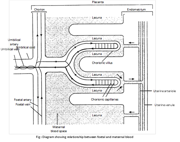
The placenta is fully formed by the end of the third month and it lasts throughout pregnancy. When complete, it is a reddish – brown disc. In the placenta, the foetal blood comes very close to the maternal blood, and this permits the exchange of materials between the two. Food (glucose, amino acids, simple proteins, lipids), water, mineral salts, vitamins, hormones, antibodies and oxygen pass from the maternal blood into the foetal blood, and foetal metabolic wastes, such as carbon dioxide and urea, also water and hormones, pass into the maternal blood. The placenta, thus, serves as the nutritive, respiratory and excretory organ of the foetus. The continuous uptake of oxygen by foetal blood is ensured by the difference in affinity for oxygen between foetal and maternal haemoglobin.
The maternal and foetal blood are not in direct contact in the placenta, because (i) the two may be incompatible; (ii) the pressure of maternal blood is far too high for the foetal blood vessels; and (iii) there must be a check on the passage of harmful materials (blood proteins, germs) into the foetal blood.
(iii) Functions
(1) Placenta helps in the nutrition of the embryo as the nutrients like amino acids, monosugars, vitamins, etc. pass from the maternal blood into foetal blood through placenta.
(2) It also helps in respiration of the embryo as \[{{O}_{2}}\] of the maternal blood and \[C{{O}_{2}}\] of the foetal blood diffuse through placenta into the foetal blood and maternal blood respectively.
(3) It also helps excretion of the embryo as nitrogenous wastes of foetal blood like urea pass into maternal blood through placenta.
(4) It also acts as an endocrine gland as it secretes certain hormones like estrogens, progesterone and human chorionic gonadorophin (HCG). HCG maintains the corpus luteum for the continued secretion of progesterone to maintain the pregnancy. At the end of gestation period, it also secretes relaxin which helps in softening of pubic symphysis and child birth. It also secrete small amounts of chorionic thyrotrophin, chorionic corticotropin and chorionic somatomammotropin.
(5) Antibodies against diphtheria, smallpox, scarlet fever, measles, etc. pass from maternal blood to foetal blood through placenta and provide passive immunity.
(6) It stores the glycogen till the formation of liver.
(7) Though the placenta acts as an effective barrier for certain toxic chemicals like histamine but certain germs like AIDS virus, syphilis bacteria, viruses of German measles, etc. intoxicants like nicotine of cigarette smoke; and addictive drugs like heroin and cocaine can pass through the placenta and cause the developmental defects.
(iv) Placenta and disease
(1) Viral and bacterial infections of placenta are known as placentitis.
(2) If the mother suffer from certain diseases like syphilis, smallpox, chickenpox, AIDS and measles; their pathogen enter into the foetus through placenta.
(3) Many drugs used medicinally may penetrate the placental barriers.
(4) Drugs such as thalidomide taken as a sedative by woman in early pregnancy (25 to 44 days) cause extensive deficiencies in the development of limbs, the alimentary canal (non perforation of the anus) and the heart in children.
(5) Nicotine from cigarette smoke crosses the placenta and stunts foetal growth.
(6) Addictive drugs such as heroin and cocaine reach the foetus, causing addiction to the new born.
(v) Classification of placenta
(a) According to the foetal membrane involved in the formation of placenta.
(1) Yolk sac placenta: In metatheria or marsupials, such as kangaroo (macropus) and opossum (Didelphys), placenta is derived from yolk sac and chorion. In metatheria, yolk sac placenta is only weakly developed so that embryonic nutrition and growth remain limited and the young is born very small and immature. To compensate the deficiency of intrauterine development, it is transferred to the abdominal pouch or marsupium and fed on milk until fully formed. In higher mammals (Eutheria) a yolk sac placenta is usually not found but in some mammals (Hedgehogs and rabbit) it may be temporarily develop in early stages.
(2) Allantoic placenta: In the majority of Eutherian, the chief organ of embryonic nutrition is the allantoic placenta consist of allantois and chorion and also called allantochorionic placenta. Outside Eutharia, a primitive allantoic placenta occurs only in perameles (bandicoot) which is a metatherian.
(3) Chorionic placenta: It occurs in primates (man and apes) and is formed only by chorion. Allantois remains small, burrows into body stalk (umbilical cord) and does not reach chorion. However, its mesoderm and blood vessels grow upto chorion whose villi enter the uterine crypts forming chorionic placenta.
(b) According to the intimacy between the foetal and maternal part.
Histologically there are six barriers are found in placenta which are as follows.
(1) Endothelium of foetal blood vessels
(2) Chorionic connective tissue
(3) Chorionic epithelium
(4) Uterine epithelium
(5) Uterine connective tissue
(6) Endothelium of maternal blood vessel
On the presence or absence of above barriers histologically placenta is divided into following types
(1) Epithelio-chorial: Most primitive and simplest type with all six placental barriers.
Examples: Odd hoofed mammals such as horse, ass, pig and lemurs.
(2) Syndesmo-chorial: Uterine epithelium absent, with five placental barriers.
Examples: Even hoofed mammals such as cow, sheep, goat, camel etc.
(3) Endothelio-chorial: Uterine epithelium and uterine connective tissues are absent, with four placental barriers.
Examples: Carnivores (dog, cat, lion, tiger etc.), Tree shrew and mole.
(4) Haemo-chorial: Uterine epithelium, uterine connective tissue and endothelium of maternal blood vessel absent, with 3 foetal layers.
Examples: Primates (man, apes and monkey).
(5) Haemo-endothelial: Foetal capillaries indirect contact with maternal blood, only one placental barrier.
Examples: Rat, guinea pig and rabbit.

(c) According to shape and distribution of villi : Depending on the shape of placenta, manner of distribution of villi, degree of connection between foetal and maternal tissues and behavior of placenta at the time of birth, the following types and subtypes of allantois placenta can be recognized.

(1) Non deciduous placenta: In most mammals villi are simple, unbranched and merely opposed without intimate contact between foetus and uterine wall. At the time of birth or parturition, villi are easily withdrawn from maternal crypts without causing any tissue damage. Thus no part of uterine tissue comes out and no bleeding occurs. Non deciduous or non-deciduate placenta has following subtypes according to the manner of distribution of villi.
(i) Diffuse: Villi remain scattered all over the surface of allantochorion. e.g. pig, horse, lemur.
(ii) Cotyledonary: Villi are arranged in separate tufts or patches called cotyledons. e.g. goat, sheep, cow, deer.
(iii) Intermediate: Villi are arranged in cotyledons as well as scattered. e.g. camel, giraffe.
(2) Deciduous placenta: Villi are complicated, branched and intimately connected. At birth, a variable amount of maternal tissue is pulled out with the shedding of blood. Deciduous or deciduate placenta is also differentiated in the following subtypes
(i) Zonary: Villi form an incomplete (e.g. racoon) or complete girdle encircling the blastocyst. e.g. cat, dog, seal.
(ii) Discoidal: Villi are restricted to a circular disc or plate on the dorsal surface of blastocyst. e.g. insectivores, bats, rodents (rat, mouse), rabbit, bear.
(iii) Metadiscoidal: Villi are at first scattered but later become restricted to one or two discs. It is monodiscoidal in man and bidiscoidal in monkeys and apes.
(3) Contra-deciduous : Foetal villi and uterine crypts are so intimately connected that even most of foetal placenta is left behind at birth to be broken and absorbed by maternal leucocytes e.g. bandicoot (perameles), mole (Talpa).
Important Tips


You need to login to perform this action.
You will be redirected in
3 sec

