Connective Tissue
Category : NEET
Connective Tissue
It connects and supports all the other tissues, the intercellular element predominating. The cellular element is usually scanty. In function this tissue may be mechanical, nutritive and defensive. It is a tissue made up of matrix (abundant intercellular substance or ground substance) and living cells that connects and support different tissues. Connective tissue was called mesenchyme by Hertwig (1893). Connective tissue is one of the most widespread tissue in the body, found in or around every organ of the body constituting about 30% of body mass and present between ectoderm and endoderm. All connective tissues in the body are formed by mesoderm.
(i) Structure: There are large intercellular spaces between the cells. Intercellular spaces are filled with large amount of extracellular materials formed of insoluble protein fibres lying in an amorphous, transparent ground substance called matrix. Ground substances is formed of mucopolysaccharides chondritin-6-sulphate of hyaluronic acid. Ageing of an animal body is associated with deterioration in its connective tissues. With advancing age, the amount of the jelly like amorphous ground substance in connective tissues of the body decreases, while the fibres become thicker and more numerous. Moreover, calcium salts get deposited in the elastin fibres of all connective tissues and particularly those of the wall of blood vessels. Consequently, the connective tissues gradually lose elasticity, resiliency and normal tone. Wrinkles in skin, poor blood supply to various tissues due to hardening of the wall of blood vessels etc., are the ultimate results of this ageing process.
(ii) Functions
(a) Attachment: Their chief function is to bind other tissues together in the organs.
(b) Storage: Certain connective tissues such as adipose tissues store fat.
(c) Support: Skeletal connective tissues like bones and cartilages provide the body with a supporting skeletal frame work.
(d) Transport: Fluid connective tissues such as blood and lymph transport various materials in the body.
(e) Defence and Scavenging: Plasma cells synthesize antibodies, viz., macrophages. Lymphocytes ingest cell debris, harmful bacteria and foreign matter. Thus these cells of connective tissues are protective in function.
(f) Shock-proof cushions: The jelly-like ground substance of connective tissues acts as shock absorber around some organs such as eye balls and kidneys.
(g) Formation of blood corpuscles: The bone marrow produces blood cells.
(h) Packing material: Areolar tissue acts as packing material in various organs.
(i) Repair: Collagen fibres of connective tissue help in repair of injured tissues.
(iii) Classification of connective tissues: It is classified into three types -
Connective tissue proper
Connective tissue proper possess soft viscous semisolid or semi-fluid matrix.
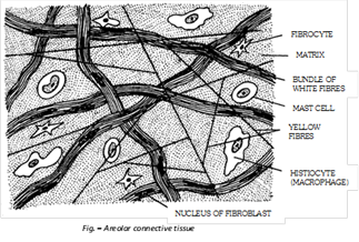
(i) Areolar Tissue: Areolar tissue is loose connective tissue, possess transparent gelatinous, highly vascular and sticky matrix which have variety of cells and fibres. It allows movement of part connected by it (Muscle and their compound). Areolar tissue mainly consist of different types of cells and fibres.
(a) Cells of areolar tissue: It has following types -
(1) Fibroblast: It is most abundant cells, produces fibres, called as fibroblasts in their young active phase and fibrocytes when old and inactive. It synthesize proteins (Collagen, elastin and reticulin). These are undifferentiated mesenchyme stem cells, capable to give rise other cells of connective tissue. Collagen and elastin are formed by fibroblasts.
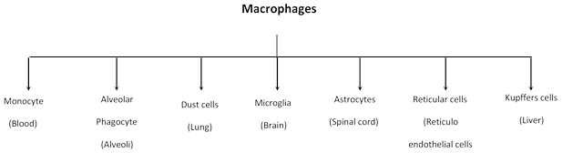
(2) Histiocytes or Macrophages or Clasmatocytes : These are polymorphic cells. These are amoeboid cells and these are main phagocytes of connective tissue. They are having most active lysosomes and phagocytise dead cells and pathogens. Some of these are called “fixed macrophage” bearing filopodia (Stellate cells) others called “wandering macrophages”. It perform “mopping up” operations. Macrophages remove the dead cells and damaged cells and clean the body so called scavenger cell. All types of macrophages take part in phagocytosis.
(3) Reticular cells: Present only in the reticular tissue and stellate in appearance. Infact they are modified fibroblast producing reticular fibres.
(4) Mast cells: Mast cells were discovered by Paul Echrlich. It is large, irregular ovoid cells found in areolar tissue. and its number increase during allergies. It produces or secretes histamine (vasodilator), serotonin (vasoconstrictor) and heparin (anticoagulant). Histamine dilate the blood vessels in allergic and inflammatory conditions. Heparin checks the clotting of blood inside the blood vessels. Serotonin act as vasoconstrictor to arrest bleeding.
(5) Lymphocytes: These are the smallest, less numerous and spherical or ovoid cells resembling lymphocytes of blood and lymph. These actively move about by pseudopodia. Their function is to form and carry antibodies. That is why, they are seen in large numbers of sites of inflammation.
(6) Plasma cells (Plasmacytes): These are usually small and rounded, superficially resembling lymphocytes but are sluggishly amoeboid and short-lived (only 2 or 3 days). These are the most potential antibody-forming cells of body presumably, mature lymphocytes (B-lymphocytes form antibody) transform into plasma cells or proliferate to form plasma cells.
(7) Fat or Adipose cells (Adipocytes or Lipocytes): A few, large and spherical cells occur in areolar tissue, singly or in clusters around small blood vessels. Each cell contains a large globule of fat surrounded by a thin peripheral layer of cytoplasm having a nucleus.
(8) Eosinophils: These cells closely resemble the eosinophilic leucocytes of blood. These probably play a phagocytic role in inflammatory and allergic reactions.
(9) Chromatophores: These are pigment cell present in specialised areas such as skin and eye. They are much branched and packed with pigment granules. They are stellate (Star like) cells, which are phagocytic in nature. They phagocytes melanin producing cells and retain melanin hence they provide colour to the skin and other organs. Melanin is black pigment which protects body from ultraviolet rays of sun.
(10) Mesenchyme cells: These are reserve undifferentiated cell which can be transformed into other types of cells when needed.
(b) Fibres of areolar tissue: These are made up with protein and non living structures of protein produced by fibroblasts and present in matrix of connective tissue and are of three types -
(1) Collagenous fibres: These are the most abundant fibrous element of areolar and other connective tissues. There are long, unbranched fibres of a soluble and shining collagen protein (tropo collagen). Some fibres and slender and straight, but most are coarse and wavy. In fact, the coarse fibres are bundles of slender fibres. Each slender fibre is, in turn, a bundle of fine fibrils, and a fibril is formed of microfibrils, which are aggregates of filamentous tropocollagen molecules. These fibres are more strengthful and provide maximum tensile strength. These are colourless and hyaline, yet called white fibres to distinguish them from yellow elastin fibres. When boiled in water, collagen (an albuminious protein) changes into gelatin. Collagen protein is the most abundant protein of the body constitutes 25% the total body protein. Collagen fibre can be stained by eosin. When collagen fibres are removed from the areolar tissue they become loose and elastic.
(2) Yellow elastin fibres: Formed of elastin protein, these fibres are less numerous, thinner, branched, anastomosing, and of a pale yellow colour. These are very elastic and remain streched due to tension in the areolar tissue, when broken in teased preparations, these coil and curl like tense wires. Elastin is probably the most resistant of all body proteins to chemical changes. Thousands of years old ‘mummies’ still have their arteries intact due to well-preserved elastin fibres. They are the orceinophilic i.e. stained by orcein.
(3) Reticulin fibres: These are delicate, freely branching and inelastic fibres of reticulin protein, found interwoven, to form networks. These are very abundant in embryos, new born babies and in healing and regenerating wounds. In areolar tissues of adults, these are mostly replaced by collagen fibres, but remain abundant in lymphoid and blood forming tissues and in the stroma of pancreas, liver etc. Chemically, reticulin is also a type of collagen. Refractive index is similar to ground substance, so without stain they can’t be seen. They are stained with AgBr and AgNO3 hence are called Argentophillic or Argyrophillic. On boiling collagen and reticular fibres both convert in glue.
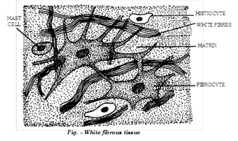
(ii) White fibrous tissue: It is modified form of areolar tissue. Only collagen fibres are present in the matrix and cells are mainly fibroblasts, present at the joints between skull bones and makes them immovable, also found in the dermis of higher mammals. It is of two types:
(a) Tendons: A tendon is non-elastic but flexible tissue consists of parallel bundles of collagenous fibres between which rows of fibroblasts are present. It joins the muscles to bones. It also form chordae tendinae which joins the cusps of atrioventricular valves of heart with the wall of ventricles.
(b) Sheath: In a sheath, the bundles of white fibres lie in a criss-cross manner. The fibroblasts are not in rows but are scattered in the areolae. The sheath form protective covering.
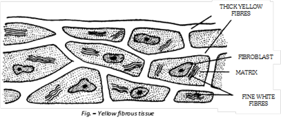
(iii) Yellow fibrous tissue: The matrix is with numerous and closely packed yellow or elastin fibres which are similar to but thicker than those of areolar connective tissue. Fibroblasts are scattered irregularly in the matrix. A few bundles of small sized white fibres are also present. It is elastic and flexible. It forms wall of blood vessels, lungs, true vocal chords, trachea, capsule of spleen and bronchioles. It also forms sheet in ligaments. Ligaments is a modified yellow elastic fibrous tissue and connects bone to bone.
(iv) Adipose tissue: It is modified form of areolar tissue made up of specialized large spherical fat cells (below the skin) or adipocytes. Mast cells and eosinophils also occur but other cell types are uncommon. Adipose tissue chiefly act as “Food reserves” or fat depots for storage and metabolism of lipids. Besides this, they also act as heat insulators and pressure, pull and push absorbers. Panniculus adiposus under the skin of human beings is partly responsible for difference in the body contours of man and women. Adipocytes are of two types:
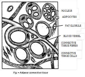
(a) Unilocular adipocyte (White adipose tissue): Common fat of body, having single large fat globule, maintain body temperature, found beneath skin subcutaneous fat panniculus adiposus, blubber of whales and elephants, hump of camel and tail of merino sheep, yellow bone marrow, around kidneys and blood vessels, mesentries, omenta and the fat bodies of frog.
(b) Multilocular adipocyte (Brown fat): Each multilocular adipocytes have several small fat globules, contain more number of mitochondria, found in rats and other rodents, polar bear, penguins, seal, walrus, in new born human babies and hibernating mammals (rats and other rodents) on oxidation it yields about 20 times more energy than ordinary fat.
(v) Reticular tissue: It is a modified form of areolar connective tissue characterized by the matrix is fluidy in nature. The matrix contains large number of stellate-shaped reticular cells, each with a number of protoplasmic processes. The protoplasmic processes of adjoining cells are interconnected to form a close network. The reticular cells secrete reticular fibres formed of a protein called reticulin. Reticular tissue is found in spleen, thymus, tonsils, lymph glands, liver, bone-marrow, lamina propria of mucosa of stomach and intestine. The reticular cells act as phagocytes and form a part of defence system of the body. Phagocytic endothelial cells of blood vessels in bone marrow and liver (kupffer cells), reticular cells of lymph nodes and spleen, macrophages of areolar tissue, dust cells of pulmonary alveoli and monocytes of blood constitute a reticulo-endothelial system. The latter is an important defence system of body, because its various components remove infective bacteria, dead cells and harmful foreign matter from blood, lymph and tissue fluid.
Important Tips
Pigmented connective tissue - Connective tissues of choroid and iris of eyes and skin dermis (corium) or black people contain large and branched (stellate) pigment cells or chromatophores (melanophores) which are laden with yellowish brown, black or blue melanin pigment granules. These are, therefore, called pigmented connective tissues. Melanin is, in fact, produced by other cells called melanocytes. Chromatophores simply phagocytes the melanin from melanocytes like macrophages.


You need to login to perform this action.
You will be redirected in
3 sec

