Epithelial Tissue
Category : NEET
Epithelial Tissue
Introduction
Body of an animal is formed of several kinds of cells. There are about 200 different types of specialized cells in the human body. The cell of one or more kinds are arranged together in a characteristic manner and cooperate to perform a specific role. Such a group of cells is called a tissue. The cell of a tissue may secrete between them a nonliving intercellular material. Thus, a tissue may be defined as a group of one or more types of cells having a similar origin and specialized for a specific function or functions along with the intercellular material.
Branch of biology dealing with the study of tissue is called histology. The term ‘tissue’ was introduced by Bichat and also known as ‘Father of histology’. Mayer coined the term ‘histology’ and the founder of histology is Marcello Malpighi. Histological study of an organ called Microscopic Anatomy. Marcello Malpighi is the father of microscopic anatomy. Hertwig introduced the term ‘mesenchyme’ for mesodermal tissue. The formation of tissues from germinal layer is called as histogenesis. The tissue classified into four main groups on the basis their location and functions, are Epithelial tissue, Connective tissue, Muscular tissue and Nervous tissue.
Epithelial Tissue
An epithelium is a tissue composed of one or more layers of cells that cover the body surface and lines its various cavities. It serves for protection, secretion and excretion. The word ‘epithelium’ was introduced by Ruysch. It was applied originally to thin skin covering the nipple (G. epi = upon, thele = nipple). They are located on the outer surfaces of organs, including the skin. They form the linings of tracts, cavities and vessels. Epithelial tissue evolved first in animal kingdom. It originate from all the three primary germ layers. e.g. Epidermis arises from ectoderm, Coelomic epithelium from the mesoderm and epithelial lining of alimentary canal from the endoderm.
(i) Structure: Cells are arranged in one or more layers, cells are compactly arranged and there is no inter cellular matrix between them. Neighbouring cells are held together by intercellular junctional complexes like desmosomes, tight junctions, interdigitations etc. the cells of lowermost - layers always rest on a non living basement membrane or basal lamina. Basement membrane is made up of no cell product of epithelial tissue. It is formed of mucopolysacharides, glycoprotein and collagen or reticular fibres. The epithelial cells are held together by small amounts of cementing substances is mainly composed of glycoprotein secreted by the cell themselves. Blood vessels are absent in the epithelial tissues. However, nerve endings may penetrate the epithelium. The free surface of cells may be smooth or may have fine hair like cilia, sterocilia and microvilli. Epithelium is subjected to continuos wear and tear and injury. Hence it posses very high capacity of renewal (mitotic cell division). The following types of modifications and junctions are found in the plasma membrane of adjacent epithelial cells to keep the cells together.

(a) Microvilli : It is simple and minute cytoplasmic processes arising from free exposed surfaces of the cell. They absorb material.
(b) Stereocilia : It is non-motile cytoplasmic processes.
(c) Cilia : It is contractile motile fibrous processes arising from basal granules.
(d) Tight junctions (Zona occludens) : At certain places the plasma membranes of adjacent cells are tightly packed or even fused together.
(e) Desmosomes : Desmosome is present in epithelial tissue. They consist of thickened area and several fine tonofibrils extending from each plasma membrane into cytoplasm of respective cells. Macula adherens is a kind of desmosome.
(f) Gap junction : At place, the adjacent cells form ion-rich gap junctions for intercellular communication and chemical exchange. These junctions probably do not provide physical support.
(g) Interdigitations : These are interwoven finger-like processes of plasma membranes of adjacent cells.
(h) Intercellular bridges : These are minute projections that arise from adjacent cell membranes. The intercellular bridges make contact with one another.

(ii) Functions : Epithelial tissues have a wide spread distribution throughout the body and serve several important functions –
(a) Protection : Generalized protection is the most important function of membranous epithelium. It is the relatively tough and impermeable epithelial covering of the skin that protects the body from mechanical
and chemical injury and also from invading bacteria and other disease causing micro-organisms.
(b) Sensation : Epithelial structures specialized for sensory functions are found in the skin, nose, eye and ear.
(c) Secretion : Glandular epithelium is specialized for secretory activity, secretory products include hormones, mucous, digestive juices and sweat.
(d) Absorption : The epithelium lining of the gut and respiratory tracts allows the absorption of nutrients from the gut.
(e) Excretion : It is the specialized epithelial lining of kidney tubules that makes the excretion and concentration of excretory products in the urine.
(f) Conduction : Ciliated epithelium moves fluid, mucous and other materials in the organs it lines.
(g) Reproduction : Germinal epithelium of the seminiferous tubules and ovaries produces spermatozoa and ova respectively.
(h) Regeneration : The ability of epithelia to regenerate quickly helps in the healing of wounds.
(i) Pigmentation : Pigmented epithelium of retina darkens the cavity of eyeball.
(j) Selective barrier : The epithelia check the absorption of harmful or unnecessary materials.
(k) Respiration : Epithelium of alveoli of the lungs brings about exchange of gases between blood and air.
(l) Exoskeleton : Epithelium also produce exoskeletal structures such as scales, feathers, hair, nail, claws, horns and hoofs.
(iii) Classification of epithelial tissue : It is mainly based on the location and functions of tissue.
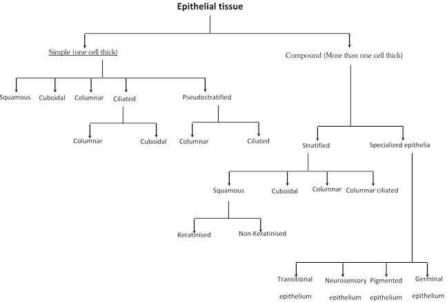
(a) Simple epithelium : It is simple in structure and basically formed by single layer cells.
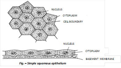
(1) Simple squamous epithelium : It is consists of only one layer of flat, scale like cells, usually polygonal cells which are closely fitted together like the tiles of a mosaic. It is also known as pavement epithelium. There is a round nucleus in the centre of cell and covers those moist places where friction causes wear and tear such as inner lining of cheeks and associated with filtration and diffusion in mammalian tissue. Blood and lymphatic vessels linings are called endothelium and surface of the pleura, pericardium and peritoneum are called mesothelium. The cells of endothelium and mesothelium become wavy and called tessellated. e.g. It forms lining of blood vessels, lymph vessel, heart, peritoneum, pleura, Bowman’s capsule, inner surface of tympanic membrane, thin segment of loop of Henle and lung alveoli.
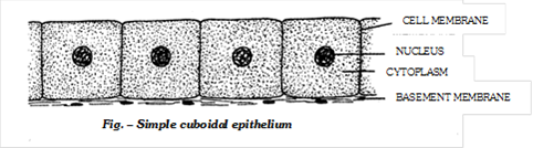
(2) Simple cuboidal epithelium : The simple cuboidal epithelium is composed of one layer of cuboidal shaped cells resting on a basement membrane. The nuclei are situated centrally. The cells of cubical epithelium often form microvilli on their free surface border called brush bordered cuboidal epithelium. e.g. the cubical epithelium is present in the small salivary and pancreatic ducts, thyroid vesicles, parts of membranous labyrinth, nephrons of kidneys, ovaries, seminiferous tubules of testes, ciliary bodies, choroid, iris of eyes, thin bronchioles and sweat gland of mammalian skin.
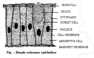
(3) Simple columnar epithelium : It consists of a single layer cells, many of which have modified structure. Three common modifications are goblet, cilia and microvilli. In the intestine plasma membranes of many columnar cells extend out in hundreds and hundreds of microscopic finger like microvilli, to increase the absorptive surface area and is called brush bordered columnar epithelium Certain cells of this epithelium contain mucous or goblet cells along with under lying supporting connective tissue is called mucous membrane. Simple columnar epithelium is present in the stomach and intestine. e.g. located inner lining of gall bladder and bile duct. It also occurs in the gastric gland, intestinal glands, pancreatic lobules, respiratory bronchioles and PCT (Proximal Convoluted Tubules).
(4) Simple ciliated epithelium : It bears numerous delicate hair like outgrowths called cilia arising from basal granules help to create a current to transport the materials. The ciliated epithelium is of two types –
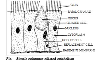
(i) Ciliated columnar epithelium : It lines respiratory tract, fallopian tubes (oviducts), ventricles of brain (ependyma), central canal of spinal cord, tympanic cavity and auditory tube (Eustachian tube).
(ii) Ciliated cuboidal epithelium : It occurs in certain parts of nephrons of the kidneys.
(5) Pseudostratified columnar epithelium : It is always consist of single layer of irregularly shaped columnar cells, touches the basement membrane. The cells are of differing heights and many are not tall enough to reach the upper surface of the epithelial sheet. Being unequal sized cells, their nuclei lie at different levels. The long cells have oval nuclei however, short cells have rounded nuclei although epithelium is one cells thick, but it gives the appearance of a stratified epithelium, hence it is called pseudostratified epithelium. Mucous secreting goblet cells are numerous and cilia are present. It is of two types –
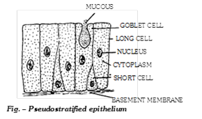
(i) Pseudostratified columnar ciliated epithelium : It is found in the lining of trachea and bronchi.
(ii) Pseudostratified columnar epithelium : It is found in certain segments of human male urethra and parotid salivary gland, vasa deferentia and epididymis.
(b) Compound epithelium : It is complexed in structure and basically formed by two or more than two layers of cells.

(1) Stratified squamous keratinised epithelium : Stratified squamous epithelium is characterized by multiple layers of cells with typical flattened squamous cells at the free or outer surface of the sheet. The presence of keratin in these cells contributes to the protective qualities of skin covering the body surface. Keratin is dead and waterproof so it protects the underlying tissues from abrasion and infection e.g. epidermis of the skin of land vertebrates.
(2) Stratified squamous non keratinised epithelium : Its free surface is moist, and the outer epithelial cells, unlike those found in the skin, do not contain keratin. This type of epithelium serves a protective function. It is found lining the oral cavity (buccal cavity), pharynx, oesophagus, anal canal, lowerpart of urethra, vocal cords, vagina, cervix (lower part of uterus) and conjunctiva of eyes.
(3) Stratified cuboidal epithelium : It is consists of two or more rows of low cuboidal-shaped cells which are arranged randomly over a basement membrane. It is found in the sweat gland ducts, larger salivary and pancreatic ducts.
(4) Stratified columnar epithelium : It is protective epithelium has multiple layers of columnar cells, only the most superficial cells are truly columnar in appearance. Epithelium of this type is rare. It is found in male urethra and in the mucous layer near the anus. It also lines mammary gland ducts and epiglottis.
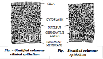
(5) Stratified columnar ciliated epithelium : It lines the larynx and upper part of the soft palate.
(c) Specialized epithelium : This type of epithelium are specialized to perform specific activity hence, specialized in structure also. They are as follows –

(i) Transitional epithelium (Urothelium) : It is often consists ten or more layers thick. It lacks germinative layer, basement membrane. Stratified transitional epithelium is typically found in the body areas such as the wall of urinary bladder, ureter and renal pelvis. It is located in all the hollow viscera subjected to stress and protects organ wall from tearing.
(ii) Neurosensory epithelium : Olfactory mucosa, called Schneiderian membrane, lining of internal nares, retina of eyes and epithelial covering of tongue containing taste buds are examples of neurosensory epithelia. These contain neurosensory cells, singly or in groups, interspersed between epithelial (supporting) cells. The sensory cells bear, at their free ends, slender “sensory hairs” to receive specific stimuli. Basely, these cells are connected, by means of synapses, with fine fibrils of sensory nerves.
(iii) Pigmented epithelium : The epithelial cells of the basal layer of retina contain pigment. Hence, this layer is often referred to as a pigmented epithelium. e.g. – Pigmented layer of retina, iris and skin.
(iv) Germinal epithelium : Specialized cuboidal cells capable of producing gametes as found in gonads. Germinal epithelium produces gametes e.g., ova (Female gametes) and sperms (Male gametes)
Glands
Glandular epithelium are specialized for secretory activity. A cell, tissue or organ which secretes a useful chemical material is known as gland. Glands are made up of cuboidal epithelial cells which are more secretory. All glands arise as folding of epithelia. The golgi body in gland cells are larger and more secretory. Most of the glands of body are merocrine types. It originate from all three germinal layers. (ecto, meso and endoderm). Liver is the largest gland of the body and lined by glandular epithelium.
(i) Types of glands
(a) Unicellular gland : It consist of unicellular gland cells which are called as goblet cells or chalice cells. They secrete mucous and found in mucosa of intestine and stomach. Mucous lubricates the food for easy peristalsis. Their life span is about 2–3 days.
(b) Multicellular gland : It consist of many cells and are generally located in underlying connective tissue e.g. gastric and intestinal glands.
(c) Exocrine gland : These are those glands which discharge their secretory products into ducts. It is also called ducted glands or glands of external secretion. e.g. Salivary glands, Mammary glands and Tear glands.
(d) Endocrine gland : It is often called ductless gland, because they discharge their secretory products (hormones) directly into the blood. e.g. Pituitary gland, thyroid, parathyroid and adrenal glands.
(e) Heterocrine gland : These are those glands which are partly endocrine and partly exocrine in function. e.g. Pancreas.
(ii) Structural classification of exocrine glands : Multicellular exocrine glands are classified by structure, using the shape of their ducts and the complexity (branching) of their ducts system as distinguishing characteristics. Shape include tubular and alveolar (Sac like). Simple exocrine glands e.g. intestinal glands, mammalian sweat glands, cutaneous glands of frog etc. have only one duct leading to surface. Compound exocrine glands have two or more ducts e.g. liver, salivary glands etc.
|
Type |
Example |
|
Simple tubular |
Intestinal glands, crypts of Lieberkuhn in ileum. |
|
Simple coiled tubular |
Sweat glands in man |
|
Simple branched tubular |
Gastric (stomach) gland, and Uterine gland. |
|
Simple alveolar |
Mucous gland in skin of frog, Poison gland of toad and seminal vesicle. |
|
Simple branched alveolar |
Sebaceous glands |
|
Compound tubular |
Brunner’s gland, bulbourethral gland and liver. |
|
Compound alveolar |
Sublingual and submandibular parotid salivary gland |
|
Compound tubulo alveolar |
Parotid salivary glands, Mammary gland and Pancreas. |
(iii) Classification of glands on the basis of their mode of secretion –
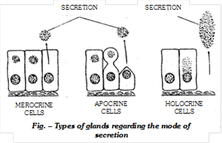
(a) Apocrine gland: Apocrine glands collect their secretory products near the apex or tip, of the cell and then release it into a duct by pinching off the distended end. This process results in some loss of cytoplasm and damage to the cell. e.g. Mammary glands. (Modified sweat gland)
(b) Holocrine gland: Holocrine glands collect their secretory products inside the cell and then rupture completely to release it. These cells self-destruct to complete their functions. e.g. Sebaceous glands. In case of rabbit sebaceous glands are found in dermis of skin. Pineal body and thymus can also be considered as holocrine gland.
(c) Merocrine gland: Merocrine glands (Eccrine or Epiccrine glands) discharge their secretory product directly through the cell or plasma membrane, without injury to the cell wall and without loss of cytoplasm. e.g. Sweat glands, exocrine region of vertebrate pancreas, salivary glands and intestinal glands etc.
(iv) Classification of glands on the basis of nature of product
(a) Mucous gland : Secret slimy mucous e.g. goblet cells, palatine gland, gland of uterus, some gastric gland and gland of colon.
(b) Serous gland : Produce watery secretion. e.g. pancreas, parotid, salivary gland, sweet gland and intestinal gland.
(c) Seromucous gland : Secrete mixed liquid. e.g. Most gastric gland, sublingual, submaxillary salivary gland.
(d) Cytogenic gland : They produce cells e.g. Testis and ovary.
Important Tips
Stereocilia present in Epididymis.


You need to login to perform this action.
You will be redirected in
3 sec

