Vascular Tissue
Category : NEET
Vascular Tissues
As the size and organizational complexity of body increased in evolution, most cells of the body got separated away from organs that receive external supplies and organs of elimination. Hence, internal transport of materials between various parts of the body became a highly specialized and important function. A vascular system, therefore, evolved in higher invertebrates and vertebrates. Blood and lymph evolved as the fluid transportation media which circulate throughout the body, carrying materials from one part to the others. These have a common fluid intercellular substance or matrix, called plasma. Several types of numerous small cells, termed corpuscles, move above or float in the plasma. There are no fibres in the plasma. Unlike other connective tissues, the plasma is not formed by the corpuscles themselves.
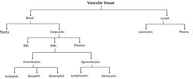
(i) Blood: In chordates, and in annelids amongst the non chordates, the blood is a red and opaque fluid of salty taste and peculiar smell. It is a little heavier than water. Its specific gravity and viscosity is 1.04 - 1.07 and 4.7 respectively. Its is 7.4 so it is slightly alkaline. In human beings, the quantity of blood is about 7% to 8% of total body weight. Thus a person, weighing about 70 kg has about 5 to 6 litres of blood, occupying about 1/13th part of the body by volume. Percentage of blood in women is slightly lower. The study of blood is called haematology. It is red coloured liquid connective tissue which originates from the mesoderm. It reaches into the various organs through the blood vessels and transports various chemical substances between different tissues. During embryonic state, the blood is mainly formed in the liver but little blood is also formed in the spleen and ribs. In adults, the blood is formed in the red bone marrow. The blood formation is called as haemopoiesis.
(ii) Plasma: It constitutes about 5% of body weight. It represents matrix of blood. Plasma is slightly alkaline and transparent. It forms 55-60% by volume of blood. Plasma contains: Water (91-92%), Solid (8-9%). Plasma solid part consists of organic (7%) and inorganic (1%) substances which are as follows:
(a) Organic constituents of plasma: Some are its own constituents, while others are those which are transported by it. All these are divisible into following categories:
(1) Plasma proteins: Protein constitute about 7% part of plasma and remain in it as colloid particles. These mainly include albumins, globulins, prothrombin and fibrinogen.
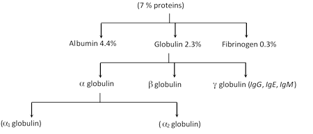
Globulins are mainly formed by plasma cells in lymphoid organs. Other plasma proteins are mainly formed in liver. These render the plasma viscous, and maintain its osmotic pressure (7.5 atmospheric) and pH. Prothrombin and Fibrinogen are essential for blood clotting. Albumins are mainly responsible for maintaining osmotic pressure in plasma and for osmoregulation in cells and tissue fluids. Globulins help in osmoregulation and transport of proteins and other substances, but most globulins are immunoglobulins, which act as antibodies, destroying harmful bacteria, virus and toxins in blood and tissue fluids. Some proteins, acting as enzymes, also occur in the plasma.
(2) Digested nutrients: These include glucose, fats, fatty acids, phospholipids, cholesterol, nucleosides, amino acids, vitamins etc. These are the supplied by the blood to all cells of body.
(3) Excretory substances: These chiefly include ammonia collected by blood from body cells and urea, uric acid, creatine, creatinine etc., collected mainly from the liver and transported to kidneys for excretion.
(4) Hormones: These are secreted and released in blood by endocrine glands.
(5) Dissolved gases: Each 100 ml. of water of blood plasma contains about 0.29 ml of O2, 5 ml. of CO2 and 0.5 ml of nitrogen dissolved in it.
(6) Defence compounds: Certain immunoglobulins or antibodies and some other substances, such as lysozyme (a polysaccharide) and properdin (a large protein) always occur in the plasma. These serve to destroy bacteria, viruses and toxic substances that may enter into the blood from outside, or from body tissues.
(7) Anticoagulant: Mast cells of connective tissues continuously release, in blood plasma, a conjugated polysaccharide, named heparin. The latter serves to prevent coagulation of blood while it is flowing in intact blood vessels.
(b) Inorganic constituents of plasma: Chloride and Bicarbonate salts of sodium are the main inorganic constituents. Traces of other salts, like phosphates, bicarbonates, sulphates and iodides of calcium, magnesium and potassium are also found. All salts constitute about 1% of plasma. These remain as ions (electrolytes) and maintain the alkalinity of plasma. A balanced quantity of salt ions in the plasma is essential for proper functioning of nervous system, muscles and other tissues.
(iii) Blood corpuscles: Blood corpuscles form 40-50% of the blood and are of three types viz. Red blood corpuscles, white blood corpuscles and platelets.
(a) Red blood corpuscles (RBC’s or Erythrocytes): These occur only in vertebrates and are the most abundant (99%) of blood corpuscles, imparting the characteristic red colour to the blood. The shape, size and structure of RBCs vary in different types of vertebrates, but their function is the same in all, namely to transport respiratory gases, especially the oxygen. (O2).
(1) RBCs of frog: Amphibian RBCs are largest amongst the vertebrates. Those of Amphiuma and Proteus are largest amongst amphibians. Those of frog measure about 35µ by 16µ (µ=1/1000 of a milimetre, i.e., 0.001 mm) and number about 4 lacs per cubic mm. of blood. These are flattened and oval, disclike, but slightly biconvex due to a large oval and centrally-placed nucleus.
(2) RBCs of mammals: Mammals have smallest RBCs amongst the vertebrates. Those of Musk deer are smallest amongst the mammals. Whereas the RBCs of other vertebrates are oval and nucleated, those of mammals are roughly circular (except those of the family camellidae - camels, llamas, dromedaries - which are oval in shape) and non-nucleated. Absence of a nucleus imparts a biconcave, disc-like shape to mammalian RBCs. During the process of their formation, mammalian RBCs lose, not only their nucleus, but also other important organelles like mitochondria, golgi bodies, centrosome, ribosomes, etc. This change in mammalian RBCs appears to be an evolutionary advancement, because it increases the surface area of RBCs and enables these to contain more heamoglobin.
(3) RBCs of human: They are about 7.4µ in diameter and its thickness is 1 to 1.5µ. It is pale yellow in colour but appear to be red in group. Surface area of all RBCs of a person totals about 1500 to 2000 times the surface area of the body itself. Erythrocyte count increases during exercise and stress, and decreases during rest, sleep, menstruation and pregnancy. Hill people have more RBCs, possibly causing their rosy cheeks. RBCs count sharply falls in anaemia and rises in polycythemia.
(4) Structure of RBCs: Each RBC is bounded by a dynamic, enzyme-containing plasma membrane. The interior has a cytoskeletal framework of a structural protein, the stromatin, and some lipids including cholesterol. The corpuscle is soft, flexible and elastic, so that it squeezes through vessels narrower than its own diameter and resumes its normal shape afterwards. In a human RBC, about 26.5 crore molecules of haemoglobin are packed in the intracellular framework. Some RBCs are probably adsorbed upon plasma membrane. Water constitutes about 60% of an RBC. The rest is solid. Haemoglobin forms about 34% of wet and 90% of dry weight of an RBC. Thus, 100 ml of normal human blood contains about 15 gm of haemoglobin on an average. An apparatus named haemoglobinometer is used to determine the haemoglobin contents of blood. Besides stromatin, lipids and haemoglobin, RBCs contain a number of enzyme systems, vitamins, salts, etc.
(5) Structure of haemoglobin: Haemoglobin is a purple coloured iron [in the form of Fe+2] containing respiratory pigment of RBCs. It consists of two parts haem (5%) and globin (95%). It is conjugated protein and made up of 4 globin chains with each attached to haem molecule by Co-ordinate bond. Globin is formed of 4 polypeptide chains \[\alpha \](141 amino acid), \[\beta \](146 amino acid), \[\gamma \](146 amino acid) and \[\delta \](146 amino acids). Each RBC contains approximately 200 to 300 million molecules of haemoglobin. One-gram haemoglobin binds 1.34 ml oxygen. Molecular formula of haemoglobin is \[{{C}_{3032}}\,{{H}_{4816}}\,{{O}_{780}}\,{{S}_{8}}\,F{{e}_{4}}\]. Amount of Hb is measured with the help of haemometer. A male has a greater amount of haemoglobin than a female. The amount of haemoglobin in normal man and woman is 14-16 gm/100 ml and 12-14 gm/100 ml respectively, while in children is slightly higher about 16.5 gm/100 ml of blood.
(6) Number of RBC: The number of RBC are counted by instrument haemocytometer. The total number of RBC per cubic mm of blood is called RBC count. RBC count is slightly lower in women than a man and number of RBC is more in people who live on mountains because there is less oxygen. RBC are absent in cockroach.
|
S.No. |
Organism |
Number of RBCs |
|
1. |
Male |
5 - 5.4 million / cubic mm of blood |
|
2. |
Female |
4.5 - 5 million / cubic mm of blood |
|
3. |
Infants |
65 - 70 lacs/ cubic mm of blood |
|
4. |
Embryo |
85 lacs/ cubic mm of blood |
|
5. |
Rabbit |
70 lacs / cubic mm of blood |
|
6. |
Frog |
4 lacs / cubic mm of blood |
(7) Life span of RBC: The life span of red blood corpuscles circulating in the blood stream varies in different animals. RBC have longest life span in blood. The mammalians RBC have short life span due to absence of nucleus, which is disappeared during development.
|
S.No. |
Organism |
Life span of RBCs |
|
1. |
Mammals and Human |
120 days or 4 months |
|
2. |
Rabbit |
80 days |
|
3. |
Frog |
100 days |
|
4. |
New born |
100 days |
(8) Function of RBCs: The major function of erythrocytes is to receive O2 of respiratory surfaces and then transport and readily deliver it to all cells of body. This important function is performed by haemoglobin which has a great ability to combine loosely and reversibly with O2 and is, hence, called “respiratory pigment”. Haemoglobin, in annelids, is dissolved in the plasma because of absence of red blood corpuscles. In mollusc and some arthropods, etc., a different respiratory pigment, haemocyanin is found dissolved in the plasma. This pigment is bluish due to presence of copper in place of iron.
(9) Formation of RBC: The process of formation of RBC is known as erythropoiesis and organ which produce RBC is called erythropoietic organs. In man erythropoiesis takes approximately 72 hrs. to complete. The process of erythropoiesis is controlled by hormone erythropoietin formed by kidney, required B12 for maturation of RBC and assisted by Fe2+. The erythrocytes are formed in liver, spleen and lymph nodes in the embryo; and in the red bone marrow in the adult. The red bone marrow is present in the cancellous bone at the extremities of long bones and between layers of compact bone in flat and irregular bones such as the cranium, vertebrae, ribs, sternum, clavicles, scapula and pelvis. The development of a mature red corpuscles takes about a week, during which the endothelial cell enlarges, divides, forms haemoglobin and finally loses its nucleus. This process is called maturation and takes place along the following lines:
Stems cells or Myeloblasts ® Proerythroblasts ® Erythroblasts ® Normoblasts ® Reticulocytes ® Erythrocytes.
Myeloblast is an amoeboid cell with fairly abundant cytoplasm and a relatively primitive, undifferentiated nucleus. Proerythroblast is larger than the myeloblast. Its nuclear network is slightly coarser and the cytoplasm is deeply basophilic due to the presence of ribonucleic acid (RNA). Erythroblasts are half the size of the proerythroblasts due to mitosis in that stage. The nucleus is checked with coarse chromatin masses. The cytoplasm is losing its RNA and is acquiring haemoglobin. Normoblasts are derived from erythroblasts by mitosis, hence they are smaller in size. The nucleus becomes progressively smaller and mitosis ceases after this stage. The cytoplasm stains strongly acidophilic due to haemoglobin. Reticulocytes are young, immature erythrocytes with a delicate network of ribonucleic acid in the cytoplasm. The nuclei of normoblasts have been lost by extrusion in this stage. Erythrocytes are mature, enucleated cells which are also called red blood corpuscles.
The normal maturation of a red blood corpuscles requires the presence of a number of different chemical substances such as vitamin B12 (cynacobalamin or erythrocyte-maturing factor) and folic acid are necessary for the development of the proerythroblast successively into erythroblast and normoblast. Vitamin B12 is present in food (mainly animal protein) but its absorption from the small intestine is dependent upon a mucopolysaccharide the intrinsic factor, secreated by the parietal cells of the gastric mucosa. Iron is necessary for the provision of haemoglobin to fill the immature erythrocytes before they can become the mature red corpuscles. Lack of iron in diet or loss of iron in bleeding causes iron-deficiency anaemia. In addition to a normal diet containing protein and iron, small amount of cobalt, copper, nicotinic acid, riboflavin and vitamin C are also essential for erythropoiesis.
The development of red blood corpuscles is controlled by a feed-back mechanism. Deficiency of oxygen following haemorrhage or because an individual, lives at high altitudes where the oxygen pressure in the atmospheric air is reduced; and a hormone called erythropoitin, secreted by the kidneys, are the two main factors that stimulate the bone marrow to increase its production of erythrocytes.
(10) Metabolism of RBCs: The mature mammalian erythrocyte lacks mitochondria; hence the cytochrome system is absent and the tricarboxylic acid cycle is not evident. The energy of the mature erythrocyte is supplied primarily by anaerobic glycolysis and the phosphogluconate pathway. RBC are created and destroyed at approximately 100 million per minute in an adult and homeostatic mechanisms operate to balance the number of cells formed against the number of cells destroyed. The excess of erythrocytes (blood) is stored in spleen, which act as a blood bank.
(11) Destruction of RBC: Aged, abnormal or damaged RBCs are phagocytosed by macrophages in the spleen and liver. Breakdowns of haemoglobin released from the RBCs yields globin and haem or iron. The globin convert to amino acids and used as an energy source or for protein synthesis. The released haem is further degraded into iron which may be stored or used immediately to produce new haemoglobin and bilirubin which is ultimately exerted in the bile.
(12) Haemolysis: Due to bursting of plasma membrane of RBCs. Its haemoglobin comes out. This process is called haemolysis. Some fat solvent and snake venom cause haemolysis. When RBCs are placed in hypotonic solution haemolysis take place. When human RBCs are placed in pure water or distilled water they will swell and burst. Some times in haemolysis, the RBCs lose their contents by diffusion and hence maintain their emptied forms intact. These are then called “shadows” or “ghosts” of RBCs.
(13) Rouleaux formation: If a drop of fresh blood is placed on a slide under coverslip. RBCs adhere together by their concave surfaces like stacks or pile coins. This is called Rouleaux formation. It occurs probably due to forces of surface tension. It may also occur temporarily in blood vessels wherever circulation becomes unduly slow for some time.
(14) ESR: It is called erythrocyte sedimentation rate. This test is measured by “Wintrobe’s tube” and “Western blotting” method. It is the rate of sinking/settling down of RBC in the plasma to form rouleaux. Man has lower ESR as compared to women and it is lowest in new born. Normal value of ESR in male is about 5 mm and in female 10 mm in first hour. A rise in ESR indicates the presence of infective/ destructive/ inflammatory diseases.
(b) White blood corpuscles (W.B.C.) or Leucocytes: They are nucleated, colourless and complete cells. They are bigger than RBC but their number is less. WBC shown least constancy in shape. The number of WBC is 8,000 to 10,000 per cubic mm. They are formed in red bone marrow, spleen, thymus and lymph nodes from myelocytes and the process is called as myelecoeisis. The life of WBC is of 15 hours to 2 days. The WBC are destroyed outside the blood vessels and the process by which the come out is called as diapedesis. An increase in the number of white blood corpuscles is called leucocytosis. More than 12,000 per cubic mm. indicates some disease. A decrease below 6000 is called leucopenia as in typhoid fever. The leucocytes are divided into two main varieties.
(1) Granular leucocytes: These cells develop in the red bone marrow from the same parent cells as the erythroblasts, i.e., myeloblast in the red bone marrow. Before entering the blood stream, they develop through promyelocyte and myelocyte stages. As these cells develop, specific granules appear in larger number and they retain their nucleus. These are granular leucocytes of roughly spherical shape, 10µ to 15µ in diameter, actively amoeboid and containing a large number of stainable granules. Their nucleus is irregular and divided into 2 to 5 interconnected lobes. Hence, these are also called polymorphonuclear leucocytes.
(i) Eosinophils (Acidophils or Oxyphils): These comprise 1% to 5% of total WBC count in blood, i.e., 70 to 300 per cu. mm. of blood. Their nucleus is distinctly bilobed with the lobes connected only by a thin strand. Their granules (lysosomes) are larger, contain important hydrolytic enzymes and stain by acid dyes like eosin. These corpuscles play important role in immunity, allergy and hypersensitivity. A rise in their number in the blood is called eosinophilia which generally occurs in parasitic worm infestations.
(ii) Basophils: These are the least numerous (only about 0.5 % to 2% of leucocytes (35 to 150 per cu. mm. of blood). Two or three lobes of their twisted, S-shaped nucleus are less distinct. Their granules (lysosomes) are larger and fewer. These stain with basic dyes like methylene blue. These corpuscles contain heparin, histamine and serotonin. Hence, these are related, but not identical, to mast cells of connective tissues.
(iii) Neutrophils (Heterophils): These are the most numerous (60 to 70% = 4000 to 5000 per cu. mm. of blood) and most active type of WBC’s. Their nucleus has 2 to 5 distinct lobes. Their granules (lysosomes) are small, but most numerous, stain with neutral dyes and contain hydrolytic enzymes capable of digesting bacteria and other pathogens. These corpuscles are actively motile and most actively phagocytic. Certain neutrophils in female mammals possess a small spherical lobe attached to their nucleus by a stalk. This lobe is called drumstick. It is formed by transformation of an X chromosome like the Barr-body of the cells of peripheral tissues in female mammals of many other species.
(2) Agranular leucocytes: They have a few non-specific or no granules in the cytoplasm and the nucleus is spherical to kidney shaped. They comprise about 25-30 % of all leucocyte and have two varieties.
(i) Lymphocytes: These are small roughly spherical (6µ to 16µ in diameter) corpuscles, comprising about 20% to 40% of the leucocytes (about 1500 to 2500 per cu. mm. of blood). These are comparatively less motile, possess a large, subspherical, central nucleus and produce antibodies. The lymphocytes are produced in the lymphatic tissue of the body which is present in the spleen, lymph nodes, thymus, tonsils and more scattered nodular masses.
Functions: Lymphocytes are concerned with the process of immunity. They produce serum globulin (b and g globulins), one of the plasma proteins, which gives rise to antibodies and antitoxins. They contribute to scar-formation after injury and thus facilitate wound-healing. Lymphocytes play an important role in the immunological reactions to tissue transplantation.
(ii) Monocytes: These comprise only about 2% to 7% of the leucocytes (i.e. 200 to 700 per cu.mm. of blood) but are the largest cells of the blood (12µ to 20µ in diameter). These have a large reniform or horse-shoe-shaped, excentric nucleus, and are actively motile and phagocytic. After entering into tissue fluid, these transform into macrophages for phagocytising invading microbes. They known as big police man of the blood. The monocytes originate from a system of primitive cells, the rectioulo-endothelial system which is found in organs such as the liver, spleen, lungs and lymphatic glands.
Functions: Their function closely resembles that of the neutrophils in that they are actively motile, phagocytic in action and will leave the blood capillaries to ingest micro-organisms and other foreign material that may be introduced into the tissue. They are the prime scavangers of cells and tissue debris. They play a vital role in removing damaged tissue and thus preparing the way for regenerative processes of the body.
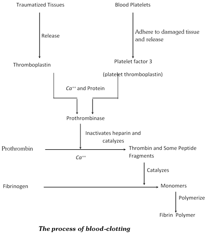
(c) Blood platelets: These are protoplasmic disks that are found in mammalian blood (lower vertebrates have spindle-shaped cells named thrombocytes). Platelets arise as detached tips of protoplasmic processes extending from the cytoplasm of giant cells, megakaryocytes, of red bone marrow. The shape is oval to round, often stellate. They are 2-3µ in diameter. The protoplasm is granular and deeply basophilic in the centre but is pale and homogenous on the periphery. There are approximately 300,000 platelets in a cubic millimetre of blood. Platelets are non-nucleated. Life span is about 10 days. Name platelet was given by Bizzazero. Agglutinated platelets are associated with blood clotting, both inside and outside of blood vessels.
(1) Coagulation or Clotting of blood: Process of formation of blood clot is also known as blood coagulation. Normal time of blood clotting is 3 to 8 minutes. Blood clotting is checked in blood vessels by presence of anticoagulant. Anticoagulant removes the cations to check the coagulation. Few important anticoagulants are heparin formed in liver and mast cells, hirudin found in leech. When an injury is caused to a blood vessel bleeding starts which is stopped by a process called blood coagulation or clotting. This process can be described under three major steps.
First step: At the site of an injury, the blood platelets disintegrate and release a phospholipid, called platelet factor-3 (Platelet thromboplastin). Injured tissues also release a lipoprotein factor called thromboplastin. These two factors combine with calcium ions (Ca++) and certain proteins of the blood plasma to form an enzyme called prothrombinase.
Second step: The prothrombinase inactivates heparin (or antiprothrombia anticoagulant) in the presence of calcium. Prothrombinase catalyzes breakdown of prothrombin (inactive plasma protein) into an active protein called thrombin and some small peptide fragments.
Third step: Thrombin acts as enzyme and first brings about depolymerization of fibrinogen ( a soluble plasma protein) into its monomers. Later thrombin stimulates repolymerization of these monomers into long insoluble fibre like polymers called fibrin. The thin, long and solid fibres of fibrin form a dense network upon the wound and trap blood corpuscles (RBCs, WBCs and platelets) to form a clot. A clot is formed at the wound in about 2 to 8 minutes after injury. The clot seals the wound and stops bleeding. Soon after the clot starts contracting and a pale yellow fluid, the serum, starts oozing out. This serum is blood plasma minus fibrinogen and blood corpuscles.
Recent theory of blood clotting is cascade theory given was Macferlane. According to this theory 13 factors are required for blood clotting.
(2) Coagulation factors
|
Factor |
Name |
Factor |
Name |
|
I |
Fibrinogen |
VIII |
Antihemophilic factor |
|
II |
Prothrombin |
IX |
Christmas factor or plasma thromboplastin component (PTC) |
|
III |
Thromboplastin |
X |
Stuart factor or Stuart-Prower factor |
|
IV |
Calcium ions |
XI |
Plasma thromboplastin antecedent (PTA) |
|
V |
Proaccelerin (Labile factor) |
XII |
Hageman factor |
|
VI |
Hypothetical factor |
XIII |
Fibrin stabilizing factor (FSF) |
|
VII |
Serum prothrombin conversion accelerator (Stable factor) |
|
|
(3) Anticoagulants
(i) Any chemical substance that prevents clotting is an anticoagulant.
(ii) Coagulation of blood in vessels is prevented during normal circulation by heparin, a quick acting anticoagulant.
(iii) Heparin inhibits conversion of prothrombin to thrombin and is used in open-heart surgery.
(iv) Vitamin K (Phylloquinone) is required for the synthesis of prothrombin necessary for blood clotting.
(v) Dicumarol acts as an antagonist for the synthesis of prothrombin necessary for blood clotting.
(vi) CPD (Citrate phosphate dextrose), ACD (Acid citrate dextrose) and EDTA (Ethylene diamino tetra acetic acid) are used by blood banks to prevent blood samples from clotting.
(vii) Blood clotting can be prevented in a test tube by adding a little oxalate or citrate (Na and K)
(viii) Oxalate or citrate react with calcium to form insoluble compound, so free calcium ions necessary for clotting are not available.
(ix) Blood is stored with an anticoagulant at 4°C At normal temperature due to potassium pump, K ions are more inside RBC than plasma. Low temperature stops the potassium pump i.e., inhibit active transport. K ions come out from RBCs resulting in ionic equilibrium.
(x) Hirudin is an anticoagulant present in the saliva of leech.
(4) Functions of blood: On basis of the above account, the general functions of blood can be briefly enumerated as follows:
(i) Transportation of materials: Blood is the fluid medium which transports different materials between various parts. It thus acts as the body’s chief “supply line”, and maintains liaison with outside environment for intake of useful materials and disposal of metabolic wastes. With the help of its haemoglobin. It takes up oxygen from external environment in respiratory organs and gives off CO2. Then, it supplies the O2 to the tissues and collects CO2 from these. In intestinal wall, it absorbs digested nutrients and distributes these to the various tissues. In return, it collects metabolic wastes from the tissues and transports these to excretory organs. It also receives hormones from endocrine glands and circulates these into all parts of the body.
(ii) Defense against infection and disease: The leucocytes of blood play the important role of defense by inactivating and destroying harmful toxins and invaders like bacteria, viruses, fungi and animal parasites.
(iii) Scavenging: Blood leucocytes phagocytes and destroy cell debris and inert foreign particles in blood and tissues. Thus, these act as “scavengers” to clean the body’s internal environment.
(iv) Control of body temperature: Blood maintains the normal temperature of body. It prevents a sharp rise or fall in temperature which may be caused in any tissue due to abnormal rate of metabolism.
(v) Healing of wounds: By coagulating at an injury, and by stimulating repairing of damaged tissues, the blood helps in rapid healing of wounds and injuries.
(vi) Homeostasis: Blood helps in the maintenance of a proper internal environment in the body by regulating the amount of salts, acids, bases and water, etc. in the tissue fluids.
(iv) Lymph: Blood, tissue fluid and lymph are almost contiguous parts of the body’s “supply line”. Infact the tissue fluid is a part of blood plasma that oozes out of arterial capillaries into intercellular substance and lymph is a part of tissue fluid. The blood plasma, tissue fluid and lymph have a basic similarity. The lymph is like the blood but, having no RBCs, it is colourless. It normally has more WBCs than the blood, and of these the lymphocytes are in large majority. It contains little of O2 , but lot of CO2 and metabolic wastes. It has the ability to coagulate like the blood. It coagulates outside the body.
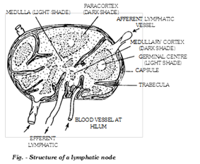
In mammals, lymph sinuses and lymph hearts are absent which are found in frog. The tissue of the mammals have lymph capillaries which join to form lymph vessels. In lymph vessel, the lymph flows from organs into the hearts. They are provided with semilunar valves to prevent reverse flow of the lymph. The lymph vessels are provided with lymph nodes which are found mainly in the head, neck, arm pits, near big blood vessels etc. The lymph nodes form lymphocytes, they clean the lymph by filtration and they form antibodies. Lymph nodes also from Payer’s patches. The lymph vessels finally open into subclavian veins. The lymph capillaries in the villi of intestine are also called as lacteals. Cisterna chyle is also called as second heart which is situated just below the diaphragm in the abdominal cavity.
Functions of lymph: The basic function of lymph is to bring back, into the vascular circulation, the cell debris, large colloid particles and the part of the blood plasma that had diffused out from arterial capillaries into the tissue fluid but has failed to return back into venous capillaries. The white corpuscles of the lymph are the same as those of the blood and have the same functions of defense and of assistance in tissue repair and healing. In intestinal wall, lymph capillaries, called lacteals, are specially meant for absorption of fats.
Comparison of blood and lymph
|
Blood |
Lymph |
|
1. Red corpuscles present. |
1. These are absent. |
|
2. White corpuscles fewer, neutrophils most numerous. |
2. White corpuscles more; lymphocytes most numerous. |
|
3. Soluble proteins more than insoluble proteins. |
3. Insoluble proteins more than soluble proteins. |
|
4. Amount of nutrients and O2 comparatively more. |
4. Amount of nutrients and O2 comparatively less. |
|
5. Amount of CO2 and metabolic wastes normal. |
5. Amount of these much more. |
Important Tips


You need to login to perform this action.
You will be redirected in
3 sec

