Implantation and Gastrulation
Category : NEET
Implantation
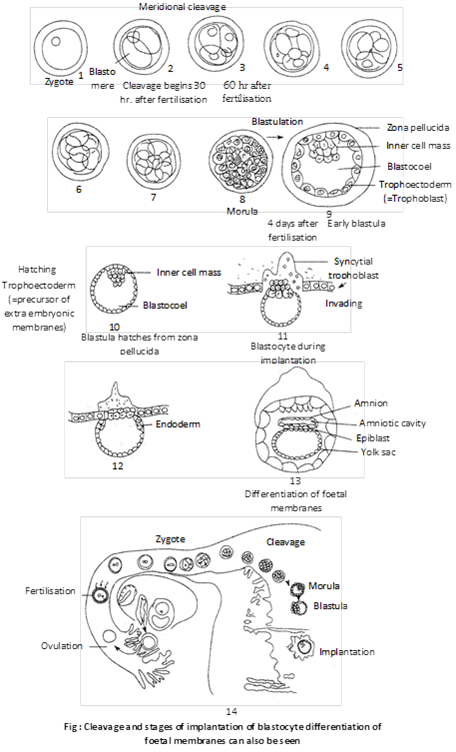
(i) Definition: The process of attachment of the blastocyst on the endometrium of the uterus is called implantation.
(ii) Period: Though the implantation may occur at any period between 6th and 10th day after the fertilization but generally it occurs on seventh day after fertilization.
(iii) Mechanism: First of all, the blastocyst is held closely against the uterine endometrial epithelium. The uterine capillaries and uterine wall in the immediate vicinity of the embryo become more permeable and a local stromal edema is developed. Soon the endometrium around the embryo shows the first sign of a decidual cell reaction (DCR) which involves:
(a) The epithelium becomes disrupted and the loosely packed fibroblast-like cells of the stoma are transformed into large rounded glycogen-filled cells.
(b) The area of contact becomes more vascular.
(c) The decidual cells form an “implantation chamber” around the embryo before the formation of a functional placenta.
(d) The tropho blast is developed from the superficial layer of the morula stage. Later, the trophoblast is lined by mesoderm to form the chorion which contributes to the placenta formation.
(e) Trophoblast of the chorion penetrates the uterine epithelium by both cytolytic and mechanical activity. The phagocytic activity of the trophoblastic cells through the decidual cells continues till it establishes intimate connection with the uterine blood vessels. The process of implantation is aided by proteolytic enzymes produced by the trophoblast. After implantation, endometrium undergoes many changes and forms decidua. It is differentiates into three parts such as : Decidua basalis present between the embryo and uterine myometrium, Decidua capsularis lies between the embryo and lumen of the uterus and Decidua parietalis is formed by the remaining part of decidua. The pattern of implantation of the blastocyst varies in different species, which are as follows
(1) Interstitial implantation: The blastocyst get burried into the endometrium e.g. human female, hedgehog, guinea pig, some bats and ape.
(2) Central implantation: The blastocyst remain the uterine cavity e.g. rabbit, cow, dog and monkey.
(3) Eccentric implantation: The blastocyst comes to lie in a uterine recess e.g. rats, mice.
(iv) Hormonal control of implantation
(a) Role of estrogens: These are a group of steroid hormones mainly secreted by follicular epithelial cells of Graafian follicle though these are also produced by adrenal cortex and placenta. These include b-estradiol, esterone, estriol etc. Out of which most important estrogen is b-estradiol. Secretion of estrogens is stimulated by FSH of anterior lobe of pituitary glands. These stimulate the uterine endometrial epithelium to enlarge, become more vascular and more glandular. The uterine glands become tortuous and cork-screw shaped. So the endometrium prepares itself for implantation. This stimulation by the estrogens on the uterus generally occurs on the 4th day of pregnancy.
(b) Progesterone: It is also a steriod hormone secreted by yellow-coloured endocrine gland, called corpus luteum, formed from empty Graafian folicle during the pregnancy. Small amount of progesterone is also secreted by adrenal cortex and placenta. Secretion of progesterone is stimulated by LH of anterior lobe of pituitary gland.
Progesterone acts on only those uterine cells which have been earlier stimulated by estrogens. Progesterone further stimulates the proliferation of endometrium of uterus and prepared, placenta formation and normal development of the foetus in the uterus.
Gastrulation
(i) Definition: Gastrulation is a dynamic process involving critical changes in the embryo such as differentiation of cells, establishment of the three primary germ layers and transformation of the single walled blastula into a double walled gastrula.
(ii) Types of gastrula movement or morphogenetic movement: The movements of cells during gastrulation is called formative or morphogenetic movements. Following types of gestural movements are found in different animals
(a) Epiboly: It involves the morphogenetic movement of prospective ectodermal (micromeres) blast meres anteroom-posteriorly to envelop the presumptive endodermal and mesodermal blast meres. It is found in telolecithal egg of frog.
(b) Emboly: It involves inward movement of prospective endodermal and chorda-mesodermal blast meres from the surface of blastula.
(c) Invagination: It involves in sinking of endodermal cells in the blastocoel to form archenteron. It is found in amphioxus.
(d) Involution: It involves the rolling in of the chorda-mesodermal blast meres inside the ectodermal cells over the lips of blastopore. It is also found in the gastrulation of frog.
(e) Convergence: It involves migration of blast meres from the outer surface towards the blastopore lips.

(f) Ingression or polyinvagination: In this, individual blastomeric migrate into the blastocoel either from only vegetal pole (called unipolar ingression e.g. Obelia; or form all sides called multipolar ingression e.g. Hydra) to form a solid gastrula called stereo gastrula.
(g) Delamination: It involves splitting off the blastoderm into two layers by the appearance of grooves resulting the formation of hypoblast. It is found in birds.
(iii) Formation of layers by gastrulation: Gastrulation includes the formation of following structures
(a) Formation of endoderm: The blast dermic vesicle enlarges and cells present on the lower surface of the embryonal knob detach by delamination from the embryonal knob. The detached cells become flat, divide increase in number and form the endoderm inside the trophoblast of the blast dermic vesicle. The embryo at this stage is tubular and encloses a hollow tube (called primitive gut or archenteron) lined by endoderm. The part of endoderm located under the embryonal knob is called embryonic endoderm which later forms embryonic gut, while the remaining part of endoderm along with trophoblast forms the yolk sac.
(b) Formation of embryonic disc and mesoderm: Meanwhile, the blastocyst continues to grow due to absorption of more and more uterine milk. The embryonal knob stretches and cells of Rubber start breaking off and dispersing. So the cells of embryonic knob from a regular layer called embryonic disc which becomes continuous with the trophoblastic. Embryonic disc is differentiated into cephalic, embryonic and caudal regions. Formation of embryonic mesoderm starts at the caudal region of the embryonic disc where cells undergo rapid proliferation and form a localized thickening of the embryonic disc and form the mesodermal layer between ectoderm and endoderm.
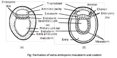
(c) Formation of ectoderm: The remaining cells of blast disc become columnar and form ectoderm.
(iv) Fate of germ layers: Each of the three germ layers gives rise to definite tissues, organs and systems of the body. Their fate in embryo and adult has been listed below
Fate of germ layer
|
Ectoderm |
Mesoderm |
Endoderm |
| Epidermis and skin derivatives |
Dermis |
Gut |
| Cutaneous gland |
Muscular tissue |
Glands of stomach and intestine |
| Nervous system (Brain + spinal cord) |
Connective tissue |
Tongue |
| Motor and optic nerve |
Endoskeleton |
Lung, trachea and bronchi |
| Eye (Retina, lens and cornea) |
Vascular system (heart and blood vessel) |
Urinary bladder |
| Conjuctiva, ciliary and iridial muscle |
Kidney |
Primordial germ cells |
| Nasal epithelium |
Gonads (Reproductive system) |
Gills |
| Internal ear (membranous labyrinth) |
Urinary and genital ducts |
Liver |
| Lateral line sense organ |
Coelom and coelomic epithelium |
Pancreas |
| Stomodaeum (mouth) |
Choroid and sclerotic coat of eye |
Thyroid gland |
| Salivary gland |
Adrenal cortex |
Parathyroid gland |
| Enamel of teeth |
Spleen |
Thymus |
| Proctodaeum | Notochord | Middle ear |
| Pituitary gland |
Parietal and visceral peritoneum |
Eustachian tube |
| Pineal body |
|
Mesoderm (Mid gut) |
| Adrenal medulla |
|
Lining of vagina and urethra |
| Hypothalamus |
|
Prostate gland |
(v) Significance of gastrulation
(a) Three primary germ, layers are formed.
(b) It marks the beginning of morphogenesis and differentiation.
(c) Metabolic activities of the cells are increased due to great morphogenetic activities of the blast meres.
Important Tips


You need to login to perform this action.
You will be redirected in
3 sec

