Nucleic Acids (DNA/RNA)
Category : NEET
Nucleic Acids (DNA/RNA)
Two types of nucleic acids are found in the cells of all living organisms. These are DNA (Deoxyribonucleic acid) and RNA (Ribonucleic acid). The nucleic acid was first isolated by Friedrich Miescher in 1868 from the nuclei of pus cells and was named nuclein. The term nuclein was given by Altman.
DNA (Deoxyribonucleic Acid)
Introduction: Term was given by Zacharis, which is found in the cells of all living organisms except plant viruses,where RNA forms the genetic material and DNA is absent. In bacteriophages and viruses there is a single molecule of DNA, which remains coiled and is enclosed in the protein coat. In bacteria, mitochondria, plastids and other prokaryotes, DNA is circular and lies naked in the cytoplasm but in eukaryotes it is found in nucleus and known as carrier of genetic information and capable of self-replication. Isolation and purification of specific DNA segment from a living organism achieved by Nirenberg H.Harries is associated with DNA-RNA hybridization technique.
(1) Chemical composition: The chemical analysis has shown that DNA is composed of three different types of compound.
(i) Sugar molecule: Represented by a pentose sugar the deoxyribose or 2-deoxyribose which derived from ribose due to the deletion of oxygen from the second carbon.
(ii) Phosphoric acid: \[{{H}_{3}}P{{O}_{4}}\] that makes DNA acidic in nature.
(iii) Nitrogeneous base: These are nitrogen containing ring compound. Which classified into two groups:
(a) Purines: Two ring compound namely as Adenine and Guanine.
(b) Pyramidine: One ring compound included Cytosine and Thymine in RNA uracil is present instead of Thymine.
Nucleosides: Nucleosides are formed by a purine or pyrimidine nitrogenous base and pentose sugar. DNA nucleosides are known as deoxyribosenucleosides.
Nucleotides: In a nucleotide, purine or pyrimidine nitrogenous base is joined by deoxyribose pentose sugar (D), which is further linked with phosphate (P) group to form nucleotides.
Composition of nucleoside and nucleotides of DNA and RNA
(D= Deoxyribose sugar, R = Ribose sugar, P = Phosphoric acid)
|
Base with its symbol |
Nucleoside |
Nucleotide |
||
|
Formula |
Name |
Formula |
Name |
|
|
DNA Adenine = A |
D - A |
Deoxyandenosine |
\[\underset{P}{\mathop{\underset{|}{\mathop{D}}\,}}\,\,-A\] |
Deoxyandenosine monophosphate or Adenine deoxyribose nucleotide |
|
Guanine = G |
D - G |
Deoxyguanine |
\[\underset{P}{\mathop{\underset{|}{\mathop{D}}\,}}\,\,-G\] |
Deoxygunine monophosphate or Guanine deoxyribose-nucleotide |
|
Thyamine = T |
D - T |
Thymidine |
\[\underset{P}{\mathop{\underset{|}{\mathop{D}}\,}}\,\,-T\] |
Thymidine monophosphate or Thymidine nucleotide |
|
Cytosine = C |
D - C |
Deoxycitidine |
\[\underset{P}{\mathop{\underset{|}{\mathop{D}}\,}}\,\,-C\] |
Deoxycytidine monophosphate or Cytosine deoxyribose nucleotide |
RNAAdenine = A |
R - A |
Adenoside |
\[\underset{P}{\mathop{\underset{|}{\mathop{R}}\,}}\,\,-A\] |
Adenosine monophosphate or Adenine ribose nucleotide |
|
Guanine = G |
R - G |
Guanosine |
\[\underset{P}{\mathop{\underset{|}{\mathop{R}}\,}}\,\,-G\] |
Guanosine monophosphate or Guanine ribose nucleotide |
|
Uracil = U |
R - U |
Uridine |
\[\underset{P}{\mathop{\underset{|}{\mathop{R}}\,}}\,\,-U\] |
Uridine monophosphate or Uracil ribose nucleotide |
|
Cytosine = C |
R - C |
Cytidine |
\[\underset{P}{\mathop{\underset{|}{\mathop{R}}\,}}\,\,-C\] |
Cytidine monophosphate or Cytosine ribose nucleotide |
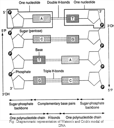
(2) Watson and Crick’s model of DNA: In 1953 Watson and Crick suggested that in a DNA molecule there are two such polynucleotide chains arranged antiparallal or is opposite directions i.e. one polynucleotide chain runs in 5’ ®3’ direction, the other in 3’® 5’ direction. It means the 3’ end of one chain lies beside the 5’ end of other in right handed manner.
(i) Important features of Watson and Crick double helical model of DNA
There are important features of DNA double helix.
(a) The double helix comprises of two polynucleotide chains.
(b) The two strands (polynucleotide chains) of double helix are anti-parallel due to phosphodiester bond.
(c) Each polynucleotide chain has a sugar-phosphate ‘backbone’ with nitrogeneous bases directed inside the helix.
(d) The nitrogenous bases of two antiparallel polynucleotide strands are linked through hydrogen bonds. There are two hydrogen bonds between A and T, and three between G and C. The hydrogen bonds are the only attractive forces between the two polynucleotides of double helix. These serve to hold the structure together.
(e) The two polynucleotides in a double helix are complementary. The sequence of nitrogenous bases in one determines the sequence of the nitrogenous bases in the other. Complementary base pairing is of fundamental importance in molecular genetics.
(f) Erwin Chargaff (1950) made quantitative analysis of DNA and proposed “base equivalence rule” starting that molar concentration of A = T & G º C or \[\frac{A+G}{C+T}\]=1&\[\frac{A+T}{G+C}\] which is constant for a species.
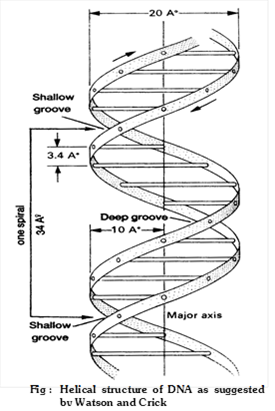
(g) Ten base pairs occur per turn of helix (abbreviated 10bp). The spacing between adjacent base pairs is 10Å. The helix is 20Å in diameter and DNA molecule found 360o in a clockwise.
(3) Forms of DNA: Five different morphological forms of DNA double helix have been described. These are A, B, C, D and Z forms. Most of these forms (except B, and Z) occur in rigidly controlled experimental conditions. Watson and crick model represents commonest form, Biotic-form (B-form or B-DNA) of DNA. Some DNA forms are inter convertible also. The differences in these DNA forms are associated with:
(i) The numbers of base pairs, present in each turn of DNA helix.
(ii) The pitch or angle between each base pair.
(iii) The helical diameter of DNA molecule.
(iv) The handedness of double helix. Which is mentioned in table.
Comparison of different types of DNA
|
Characters |
A-DNA |
B-DNA |
C-DNA |
D-DNA |
Z-DNA |
|
Base pair per turn of the helix |
11 |
10 |
9.33 |
8 |
12 |
|
Tilt of pairs (\[\gamma \]) base |
\[20.2{}^\circ \] |
\[6.3{}^\circ \] |
\[-7.8{}^\circ \] |
\[-16.7{}^\circ \] |
\[7\text{ }{\AA}\] |
|
Axial rise (h) |
\[2.56\text{ }{\AA}\] |
\[3.37\text{ }{\AA}\] |
\[3.32\text{ }{\AA}\] |
\[3.03\text{ }{\AA}\] |
\[3.7\text{ }{\AA}\] |
|
Helical diameter |
\[23\text{ }{\AA}\] |
\[20\text{ }{\AA}\] |
\[19\text{ }{\AA}\] |
- |
\[18\text{ }{\AA}\] |
|
Handedness of the double helix |
Right handed |
Right handed |
Right handed |
Right handed |
Left handed |
(4) Characteristics of DNA
(i) Denaturation or melting: The phenomenon of separating of two strand of DNA molecule by breaking of hydrogen bond at the temp.\[90{}^\circ C\].
(ii) Renaturation or annealing: Separated strands reunite to form double helix molecule of DNA by cooling at the room temp. i.e. \[25{}^\circ C\].
These properties help to form hybrid from different DNA or with RNA.
(5) Evidences of DNA as the genetic material: The following experiments conducted by the molecular biologists provide direct evidences of DNA being the genetic material bacterial transformation, bacterial recombination and bacteriophage infection.
(i) Bacterial transformation or Griffith's Experiments: Griffith (1928) injected into mice with virulent and smooth (S-type, smooth colony with mucilage) form of Diplococcus pneumoniae. The mice died due to pneumonia. No death occurred when mice were injected with nonvirulent or rough (R-type, irregular colony without mucilage) form or heat- killed virulent form. However, in a combination of heat killed S-type and live R-type bacteria, death occurred in some mice. Autopsy of dead mice showed that they possessed S-type living bacteria, which could have been produced only by transformation of R-type bacteria. The transforming chemical was found out by O.T.Avery, C.M. Mc. leod and M. Mc. Carty (1944). They fractionated heat-killed S-type bacteria into DNA, carbohydrate and protein fractions. DNA was divided into two parts, one with DNAase and the other without it. Each component was added to different cultures of R-type bacteria. Transformation was found only in that culture which was provided with intact DNA of S-type. Therefore, the trait of virulence is present in DNA. Transformation involves transfer of a part of DNA from surrounding medium or dead bacteria (donor) to living bacteria (recipient) to form a recombinant.
(ii) Evidence from genetic recombination in bacteria or bacterial conjugation: Lederberg and Tatum (1946) discovered the genetic recombination in bacteria from two different strains through the process of conjugation. Bacterium Escherichia coli can grow in minimal culture medium containing minerals and sugar only. It can synthesize all the necessary vitamins from these raw materials. But its two mutant strains were found to lack the ability to synthesize some of the vitamins necessary for growth. These could not grow in the minimal medium till the particular vitamins were not supplied in the culture medium.
(a) Mutant strain A: It (used as male strain) had the genetic composition \[Me{{t}^{}},\text{ }Bi{{o}^{}},\text{ }Th{{r}^{+}},\text{ }Le{{u}^{+}},\text{ }Th{{i}^{+}}\]. It lacks the ability to manufacture vitamins methionine and biotin and can grow only in a culture medium which contains these vitamins in addition to sugar and minerals.
(b) Mutant strain B: It (used as female strain or recipient) has a genetic composition \[M{{e}^{++}},\text{ }Bi{{o}^{+}},\text{ }Th{{r}^{}},\text{ }Le{{u}^{}},~Th{{i}^{}}\]. It lacks the ability to manufacture threonine, leucine and thionine and can grow only when these vitamins are added to the growing medium.
These two strains of E.coli are, therefore, unable to grow in the minimal culture medium, when grow separately. But when a mixture of these two strains was allowed to grow in the same medium a number of colonies were formed. This indicates that the portion of donor DNA containing information to manufacture threonine, leucine and thionine had been transferred and incorporated in the recipient’s genotype during conjugation.
This experiment of Lederberg and Tatum shown that the conjugation results in the transfer of genetic material DNA from one bacterium to other. During conjugation a cytoplasmic bridge is formed between two conjugating bacteria.
(iii) Evidence from bacteriophage infection: Hershey and Chase (1952) conducted their experiment on \[{{T}_{2}}\] bacteriophage, which attacks on E.coli bacterium. The phage particles were prepared by using radioisotopes of \[^{\mathbf{35}}\mathbf{S}\] and \[^{\mathbf{32}}\mathbf{P}\] in the following steps.
(a) Few bacteriophages were grown in bacteria containing \[^{\mathbf{35}}\mathbf{S}\]. Which was incorporated into the cystein and methionine amino acids of proteins and thus these amino acids with \[^{\mathbf{35}}\mathbf{S}\] formed the proteins of phage.
(b) Some other bacteriophages were grown in bacteria having \[^{32}P\]. Which was restricted to DNA of phage particles. These two radioactive phage preparations (one with radioactive proteins and another with radioactive DNA) were allowed to infect the culture of E.coli. The protein coats were separated from the bacterial cell walls by shaking and centrifugation.
The heavier infected bacterial cells during centrifugation pelleted to bottom. The supernatant had the lighter phage particles and other components that failed to infect bacteria. It was observed that bacteriophages with radioactive DNA gave rise to radioactive pellets with \[^{32}P\] in DNA. However in the phage particles with radioactive protein (with\[^{35}S\]) the bacterial pellets have almost nil radioactivity indicating that proteins have failed to migrate into bacterial cell. So, it can be safely concluded that during infection by bacteriophage \[{{T}_{2}}\], it was DNA, which entered the bacteria. It was followed by an eclipse period during which phage DNA replicates numerous times within the bacterial cell. Towards the end of eclipse period phage DNA directs the production of protein coats assembly of newly formed phage particles. Lysozyme (an enzyme) brings about the lysis of host cell and release, the newly formed bacteriophages. The above experiment clearly suggests that it is phage DNA and not protein, which contains the genetic information for the production of new bacteriophages. However, in some plant viruses (like TMV), RNA acts as hereditary material (being DNA absent).
(6) DNA replication : Watson and Crick suggested a very simple mechanism of DNA replication or DNA transcription on the basis of its double helical structure. During replication the weak hydrogen bonds between the nitrogenous bases of the nucleotides separate so that the two polynucleotide chains of DNA also separate and uncoil. The chains thus separated are complementary to one another. Because of the specificity of base pairing, each nucleotide of separated chains attracts it complementary nucleotide from the cell cytoplasm. Once the nucleotides are attached by their hydrogen bonds, their sugar radicals unite through their phosphate components, completing the formation of a new polynucleotide chain.
The method of DNA replication is semi-discontinuous and described as semi-conservative method, because each daughter DNA molecule is a hybrid conserving one parental polynucleotide chain and the other one newly synthesized strand. DNA replication occur in S-phage in cell cycle.
(i) Mechanism of DNA replication
The entire process of DNA replication involves following steps in E.coli.
(a) Recognition of the initiation point: First, DNA helix unwind by the enzyme “Helicase” which use the energy of ATP and replication of DNA begin at a specific point, called initiation point or origin where replication fork begins.
(b) Unwinding of DNA: The unwinding proteins bind to the nicked strand of the duplex and separate the two strands at DNA duplex. Topoisomerase (Gyrase is a type of topoisomerase in E.coli) helps in unwinding of DNA.
(c) Single stranded binding protein (SSB): Which remain DNA in single stranded position and also known as helix destabilising protein (HDP).
(d) RNA Priming: The DNA directed RNA polymerase now synthesizes the primer strands of RNA (RNA primer). The priming RNA strands are complementary to the two strands of DNA and are formed of 50 to 100 nucleotides.
(e) Formation of DNA on RNA primers: The new strands of DNA are formed in the 5¢®3¢ direction from the 3¢®5¢ template DNA by the addition of deoxyribonucleotides to the 3¢ end of primer RNA.
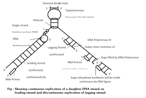
Addition of nucleotide is done by DNA polymerase III. The leading strand of DNA is synthesized continuously in 5¢®3¢ direction as one piece. The lagging strand of DNA is synthesized discontinuously in its opposite direction in short segments. These segments are called Okazaki fragments.
(f) Excision of RNA primers: Once a small segment of an okazaki fragment has been formed. The RNA primers are removed from the 5¢ by the action of 5¢®3¢ exonuclease activity of DNA polymerase I.
(g) Joining of okazaki fragments: The gaps left between Okazaki fragments are filled with complimentary deoxyribonucleotide residues by DNA polymerase-I. Finally, the adjacent 5¢ and 3¢ ends are joined by DNA ligase.
(ii) DNA polymerase enzymes : There are three DNA polymerase enzymes that participate in the process of DNA replication.
(a) DNA polymerase-I: This enzyme has been studied in E. coli in detail. It possesses a sulphydryl group, single interchain disulphide and one zinc molecule at the active site. DNA polymerase-I was discovered by Kornberg and his colleagues in 1955. It was considered to carry out DNA replication and also participates in the repair and proof reading of DNA by catalyzing the addition of mononucleotide units (the deoxyribonucleotide residues) to the free 3¢ -hydroxyl end of DNA chain. A pure DNA polymerase-I can add about 1,000 nucleotide residues per minute per molecule and catalyses 5¢ ®3¢ exonuclease activity and removes nucleotide residues of primer RNA at 3¢.
(b) DNA polymerase-II: The biological role of polymerase II is not yet known.
(c) DNA polymerase-III: This enzyme was discovered by T. Kornberg and M.L. Gefter (1972). It is the most active enzyme and responsible for DNA chain elongation.
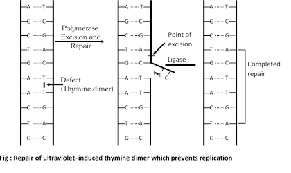
(iii) DNA repair: When DNA damaged by mutagen, a system is activate to repair damage DNA. Say for example UV light induced thymidine dimers in DNA and repair mechanism of that DNA called photo reactivation. Many enzyme involved in repair mechanism in which endonuclease (Chemical knives) cut the defective part of DNA then gap is filled with DNA polymerase I and finally DNA ligase seals that repaired part.
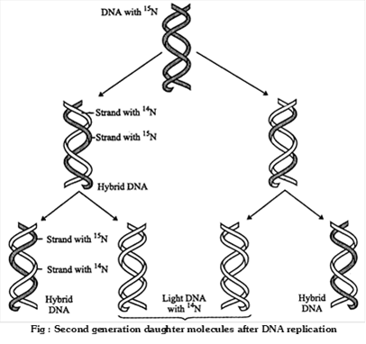
(iv) Evidence in support of semiconservative mode of DNA replication (Meselson and Stahl’s experiment): Meselson and Stahl (1958) cultured (Escherichia coli) bacteria in a culture medium containing \[{{\mathbf{N}}^{\mathbf{15}}}\]were isotopes of nitrogen. After these had replicated for a few generations in that medium both the strands of their DNA contained \[{{\mathbf{N}}^{\mathbf{15}}}\] as constituents of purines and pyrimidines. When these bacteria with \[{{\mathbf{N}}^{\mathbf{15}}}\] were transferred in cultural medium containing \[{{\mathbf{N}}^{\mathbf{14}}}\], it was found that DNA separated from fresh generation of bacteria possesses one strand heavier than the other. The heavier strand represents the parental strand and lighter one is the new one synthesized from the culture indicating semiconservative mode of DNA replication. circular form of replication on as characteristic of prokaryotes is theta replication discovered by J. Cairns.
Important Tips
Central Dogma
Central dogma of molecular biology proposes a unidirectional or one way flow of information from DNA to RNA (transcription) and from RNA to protein (translation). The concept was given by Watson and Crick.
\[\text{DNA}\xrightarrow{\text{Transcription}}\text{mRNA}\ \xrightarrow{\text{Translation}}\ \text{Protein}\]
As mentioned above the first step of central dogma is transcription (synthesis of mRNA from DNA), but in case of reverse transcription DNA is synthesizes from RNA in retrovirus. That concept is given by Temin and Baltimore in Rous sarcoma virus, also known as teminism and enzyme catalyze this reaction is reverse transcriptase or RNA dependent DNA polymerase.
\[\text{DNA}\underset{\xleftarrow[\text{Reverse}\,\text{Transcription}]{}}{\mathop{\xrightarrow{\ \ \ \ \text{Transcription}\ \ \ \ \ }}}\,\text{RNA}\xrightarrow{{}}\text{Protein}\]
Transcription
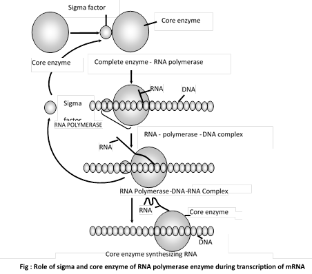
Formation of mRNA from DNA is called as Transcription. It is heterocatalytic function of DNA. Template of DNA called sense strand (Master Strand) is involved. The segment of DNA involved in transcriptions is cistron, which have a promoter region where initiation is start and terminator region where transcription ends. Enzyme involved in transcription is RNA polymerase-II. Which consist five polypeptide \[\alpha ,\beta ,{{\beta }^{'}},\omega \] (constitute core enzyme) and \[\sigma \](sigma factor). Sigma \[(\sigma )\] factor recognise promoter site while remaining core enzyme takes part in chain elongation. After transcription, DNA molecule reassociates to form its original structure. In eukaryotes hn RNA (heterogenous nuclear RNA) which consist exon (coded region) and introns (non coded region or intervening sequences) formed in nucleus and diffuse in cytoplasm is also known as split gene which goes to transcription changes for removing the introns and later formed mRNA.
It consist three phenomenon: -
(1) Initiation: Initiation start with help of \[\sigma \](sigma) factor of RNA polymerase enzyme. At the cap region which have 7 methyl guanosine residue at the 5¢.
(2) Elongation: Elongation is done by core enzyme, which moves along the sense strand.
(3) Termination: In prokaryotes termination is done by rho (\[\rho \]) factor while in eukaryotes poly A tail is responsible for termination at the 3¢.
Important Tips


You need to login to perform this action.
You will be redirected in
3 sec

