Nervous Tissue
Category : NEET
Nervous Tissue
A most complex tissue in the body, composed of densely packed interconnected nerve cells called neurons (as many as 1010 in the human brain). It specialized in communication between the various parts of the body and in integration of their activities. Nervous tissue is ectodermal (from neural plate) in origin. It forms the nervous system of the body which controls and coordinates the body functions. Nerve cells (neurons) are specialized to receive the external and internal stimuli. A stimulus of adequate strength (threshold stimulus) causes the depolarization or reversal of polarity of the neuron locally and initiates a nerve impulse. The neurons are capable of conducting this depolarization as a wave along their length in a particular direction either to other nerve cells or to effectors like muscles and glands which give the response. There response may be in the form of muscle contraction or glandular secretion. Therefore, excitability and conductivity two fundamental properties of nervous tissues. There is no intercellular matrix between neurons. These have permanently lost the power of division as have no centriole and have minimum power of regeneration. So these cannot be cultured in vitro. Irritability is the main function of nervous tissue.
(i) Composition of Nervous Tissue: Nervous tissue is formed of four types of cells:
(a) Neurons (nerve cells)
(b) Neuroglia
(c) Ependymal cells
(d) Neuro-secretory cells
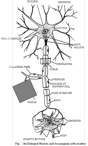
(a) Structure of neurons: A neuron is a nerve cell with all its branches. Neuron is formed from neuroblast. It is the structural and functional unit of nervous system. It is the longest cell of the body.
(1) Cyton: It is also called perikaryon or soma or cell body. Its granular cytoplasm is called neuroplasm which has following structures :
(i) A large, spherical, centrally placed nucleus with a single nucleolus.
(ii) Numerous fine threads called neurofibrils for the conduction of nerve impulses.
(iii) A number of small, basophilic granules called Nissl’s granules formed of rough endoplasmic reticulum with ribosomes and are sites of protein synthesis.
(iv) Neuroplasm has large number of mitochondria to provide high energy for impulse conduction.
(v) Neuroplasm may have melanophores with melanin pigment and lipochromes with orange or yellow pigment.
(vi) A mature neuron has no centriole, so it cannot divide.
(vii) A “Barr body” is often seen abutting against the inner surface of nuclear membrane of cytons in females. This has been proved to be a transformed ‘X’ chromosome.
(viii) Certain neurons having flask-shaped cytons and called purkinje cells, occur in the cerebellum of the brain.
(2) Neuron processes: The processes of neurons, called neurites, extend varying distances from the cyton and are of two types - dendrites or dendrons and an axon or axis cylinder (neuraxon).
(i) Dendron: These are several short, tapering much branched processes. The dendrites contain neurofibrils, neurotubules, Nissl’s granules and mitochondria. They conduct nerve impulse towards the cell body.
(ii) Axon: This is a single very long, cylindrical process of uniform diameter. It arises from a conical projection, the axon hillock, of the cyton. The axon contains neurofibrils and neurotubules but lacks Nissl’s granules. The axon is therefore dependent on the cell body for supply of proteins. The cell membrane of axon is called axolemma and its cytoplasm is called axoplasm. The axon conducts impulses away from the cell body. It may give off lateral branches termed collateral fibres. The latter arise from a node at right angle. Axon is usually branched only terminally into slender branches called telodendria. The latter have knobbed ends called endbulbs or axon terminals or buttons or synaptic knobs or end plates. The synaptic knobs contain mitochondria and secretory vesicles.
Differences between Axon and Dendron
|
Characters |
Axon |
Dendron |
|
1. Number |
Always single |
May be one or more in number |
|
2. Structure |
Formed of neuroplasm with only neurofibrils but no Nissl?s bodies. |
Formed of neuroplasm with both neurofibrils and Nissl?s bodies |
|
3. Size |
Long sized processes |
Small sized processes |
|
4. Direction of new impulses |
Always away from the cell body |
Always towards the cell body |
|
5. Nature |
Efferent |
Afferent |
|
6. Branching |
Generally absent |
Generally present |
(b) Neuroglia or Glia cells: Neuroglia consists of the supporting and packing cells found in the brain, spinal chord and ganglia. These are non nervous cells. These are ten times more numerous than neurons. In some parts of body the neuroglial cells are called by certain other name such as muller cells in retina, pituicytes in posterior pituitary gland and satellite cells in ganglia.
(1) Types: The neuroglia cells are of three types -
(i) Astrocytes: These are large sized and star-shaped cells with numerous processes.
(ii) Oligodendrocytes: These have a few branched processes which resemble the dendrons of the neurons.
(iii) Microglial cells: These are small sized and spindle-shaped. The microglia cells act as the defensive phagocytes in central nervous system. They arise from the monocytes.
Differences between Neurons and Neuroglia
|
Neurons (Nerve cells) |
Neuroglia (Glial cells) |
|
1. Have a relatively small cell body and long processes. |
1. Have a relatively large cell body and short processes. |
|
2. Processes arise from the two opposite ends of the cell body. |
2. Processes arise from nearly all over the cell body. |
|
3. Processes are of 2 types : short dendrons and along axons. |
3. Processes are all alike. |
|
4. Neurons occur end to end in chains. |
4. Glial cells are aggregated in masses. |
|
5. Neurons set up and conduct nerve impulses |
5. Glial cells form a supporting and packing tissue that insulates the neurons. Some (microglia cells) are phagocytic. |
|
6. All neurons arise from the ectoderm. |
6. Most glial cells arise from the ectoderm, microglia cells arise from the monocytes. |
|
7. Neurons form synapses. |
7. Glial cells do not form synapses. |
(2) Functions:
(i) These are capable of division and help in wear and tear of the central nervous system.
(ii) These insulate the adjoining neurons and prevent the lateral transmission of impulses.
(iii) These provide nutrition to the neurons.
(iv) These act as phagocytes and eat up the microbes.
(v) These help in memory processes.
(vi) They acts as Blood brain barrier (BBB) i.e. they inhibit contact between neuron and blood, along with endothelium of capillary. The exchange of material between blood and neuron is always through these neuroglial cells i.e., they are mediator.
(c) Ependymal cells: These are cuboidal and ciliated epithelial cells which lines the cavities of brain (ventricles) and spinal chord (central canal). These form an epithelium called ependyma.
(d) Neurosecretory cells: These are special type of neurons of the hypothalamus of brain. These are endocrine in function and secrete neurohormones which are carried by the blood of hypophyseal portal system to anterior lobe of pituitary gland and stimulate the secretion of their trophic hormones e.g., TSH, STH, FSH, LH, ACTH, etc.
(ii) Types of neurons: Neurons are divided into different categories on different basis.
(a) On the basis of functions: Neurons are divided into three categories :
(1) Sensory (afferent) neurons: These are found in sense organs. Their dendrons receive the nerve impulse from the nerve process of the receptor cell while their axon forms the synapse with dendron of the next neuron. These may be naked or encapsulated e.g. olfactoreceptors and gustatoreceptors.
(2) Internuncial neurons: These are located in the dorsal horn of the spinal cord. Their dendrons form the synapse with the axon of sensory neuron while their axon forms the synapse with the dendron of the motor neuron. These are called association neurons (when their axon synapses with the dendron of motor neuron of same side) or commissural neuron (when their axon synapses with the dendron of motor neuron of opposite side).
(3) Motor (efferent) neurons: These are always present in the ventral horn of the spinal cord. Their axon ends into the muscle fibres or glands cells. These conduct the nerve impulses to the effector organs which respond to the stimuli.
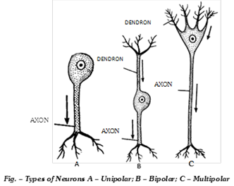
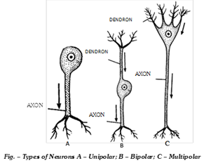
(b) On the basis of number of nerve processes: Neurons are of three types -
(1) Unipolar neurons: In these neurons, only one nerve process arises from the cyton which acts as axon but there is no dendron. These are found only in early embryos. The unipolar neuron of the adult gives rise to a single nerve process, which immediately divides into a dendron and an axon. Such unipolar neurons are called pseudo-unipolar neurons. These are found in the dorsal root ganglia of spinal nerves and in the roots of V, IX and X cranial nerves.
(2) Bipolar neurons: In these neurons, the cyton gives rise to two nerve processes out of which one acts as an axon while other acts as a dendron. These are found in the olfactory epithelium of nasal chamber and retina of eye. These may be isopolar or heteropolar (dendrons being irregularly branched).
(3) Multipolar neurons: In these neurons, the cyton gives rise to several nerve processes out of which one acts as an axon while remaining nerve processes act as dendrons. These are found in the central nervous system and the ganglia of autonomic nervous system.
(iii) Nerve fibres: Axon or dendron of a nerve cell covered with one or two sheath is termed as nerve fibre. The nerve fibres are of two types - medullated or myelinated and non medullated or non myelinated regarding their structure.
(a) Medullated nerve fibres: A medullated nerve fibre typically consists of a central core, the axis cylinder, or neuraxis, surrounded by two sheaths : inner thick medullary sheath and outer thin neurilemma.
(1) Axis cylinder: The axis cylinder is simply the axon or dendron of a nerve cell. It contains longitudinal neurofibrils and mitochondria in its neuroplasm, called axoplasm, limited by cell membrane termed axolemma. It is the axolemma that conducts the nerve impulses.
(2) Medullary sheath: The medullary sheath is composed of a shinning, white, fatty substance called myelin. This sheath perhaps serves as an insulating layer, preventing loss of energy of the nerve impulse during its passage along the fibre. The medullary sheath is continous around the fibres in the central nervous system, but in the fibres of the peripheral nerves it is absent at certain points known as the Node of Ranvier. The part of a nerve fibre between two successive nodes is termed the internode.
(3) Neurilemma: The neurilemma consists of tubular sheath cells (Schwann’s cells) placed end to end. The neurilemma is continuous over the Nodes of Ranvier. The function of the Schwann’s cells is to produce the myelin sheath around the neuraxis. Outside neurilemma is a thin layer of connective tissue. It is called endoneurium. It keeps the nerve fibre held to the others in a nerve. The medullated nerve fibres within the brain and spinal chord lack neurilemma. Instead, they have an incomplete covering of neuroglia cells, which probably produce the myelin sheath. Neurilemma present around the peripheral nerve fibres enables them to regenrate after injury. Nerve fibres in the brain and spinal chord do not regenrate after injury due to lack of neurilemma. The medullated nerve fibres occur in the white matter of the brain and spinal chord and in the cranial and spinal nerves.
(b) Non medullated nerve fibres: A non medullated nerve fibre consists of an axis cylinder enclosed by neurilemma and connective tissue. These fibres appears grey in colour in the fresh state. The non-medullated nerve fibres occur in the autonomic nerves.
Difference between medullated and non-medulated nerve fibre
|
Characters |
Medullated nerve fibres |
Non-medullated nerve fibres |
|
1. Occurrence |
Found in white matter of brain, spinal cord, cranial and spinal nerves |
Found in grey matter of brain and spinal cord, and in autonomic nervous systems. |
|
2. Sheaths. |
Neuraxis covered by inner medullary sheath and outer neurilemma |
Neuraxis covered by only neurilemma. Medullary sheath is absent |
|
3. Nodes of Ranvier and internodes |
Present |
Absent |
|
4. Diameter |
More |
Less |
|
5. Colour |
White |
Grey |
|
6. Speed of conduction of nerve impulses. |
Faster due to saltatory conduction of nerve impulses |
Slower |
|
7. Collateral branches |
Present |
Absent |
(iv) Nerves
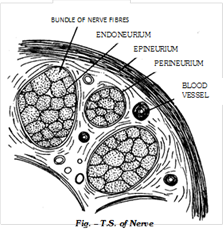
(a) Structure: The nerves are thread like structures extending between the central nervous system and the receptor of effector organs of the body. These conduct the nerve impulses to and from the central nervous system.
Each nerve is formed of several bundles of nerve fibres, called fasciculi. Each nerve fibre of the bundle is covered by a thin sheath of connective tissue called endoneurium, while each fasciculus is enclosed by another sheath of white fibrous connective tissue called perineurium. All the fasciculi are held together by the connective tissue and are enclosed by a thick coat of white fibrous connective tissue called epineurium. On average, a nerve contains about twice as many unmyelinated fibres as myelinated fibres.
(b) Types of nerves: The nerves are of three types according to the nature of the nerve fibres they are composed of
(1) Sensory or afferent nerves: The nerves with sensory fibres are called sensory nerves. Example - Olfactory, optic and auditory nerves.
(b) Motor or efferent nerves: The nerves having efferent fibres are termed motor nerves. Example - Oculomotor, Pathetic and abducens nerves.
(c) Mixed nerves: Some nerves have both afferent and efferent fibres. These are known as mixed nerves. Example - Trigeminal, facial, glossopharyngeal and vagus nerves.
Important Tips


You need to login to perform this action.
You will be redirected in
3 sec

