Tissue (General)
Category : NEET
Tissue (General)
Introduction
Plant tissue is a collection of similar cells performing an organized function for the plant. Each plant tissue is specialized for a unique purpose, and can be combined with other tissues to create organs such as leaves, flowers, stems and roots. The following is a brief outline of plant tissues, and their functions within the plant.
Meristematic tissues or Meristems
The word “Meristem” originated from “Meristos” (Greek = continuous division) and the term meristem was introduced by Nageli (1858). A group of cells which are much active and capable of showing continuous divisions and redivisions, is called as meristematic tissue. The various characteristic features of the meristems are discussed below :
(1) Types of meristems : The meristems may be classified on the basis of their mode of origin, position or function :
(i) According to origin and development : On the basis of origin, meristematic tissues are of three types :
(a) Promeristem or Primordial meristem : The promeristem originates from embryo and, therefore, called primordial or embryonic meristem. It is present in the regions where an organ or a part of plant body is initiated. A group of initial cells that lay down the foundation of an organ or a plant part, is called promeristem. This group consists of a limited amount of cells, which divide repeatedly to give rise primary meristem. It occupies a small area at the tips of stem and root. The promeristem gives rise to all other meristems including the primary meristem.
(b) Primary meristem : A primary meristem originates from promeristem and retains its meristematic activity. It is located in the apices of roots, stems and the leaf primordia. Primary meristem gives rise to the primary permanent tissue.
(c) Secondary Meristem : They always arise in permanent tissues and have no typical promeristem. Some living permanent cells may regain the meristematic nature. This process in which permanent tissue regains meristematic nature is called dedifferentiation. The secondary meristems are so called because they originate from permanent cells. The phellogen or cork cambium arising from epidermis, cortex or other cells during secondary growth, is an important example of secondary meristem. The secondary meristems produce secondary tissues in the plant body and add new cells for effective protection and repair.
(ii) According to position : On the basis of their position in the plant body meristems are classified into three categories :
(a) Apical meristem : This meristem is located at the growing apices of main and lateral shoots and roots. These cells are responsible for linear growth of an organ. The initiating cells may be single or in groups. Solitary initial cells are known as apical cells whereas those occurring in groups are called apical initials. Solitary apical cells occur in ferns and other Pteridophytes while apical initials are found in other vascular plants. The apical initials may occur in one or more tiers. Position of apical cells may either be strictly terminal or terminal and subterminal.
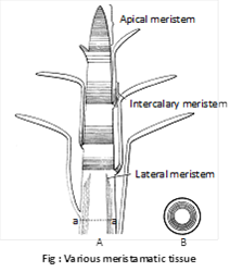
(b) Intercalary meristem : These are the portions of apical meristems which are separated from the apex during the growth of axis and formation of permanent tissues. It is present mostly at the base of node (e.g., Mentha viridis-Mint), base of internode (e.g., stem of many monocots viz., Wheat, Grasses, Pteridophyts like Equisetum) or at the base of the leaf (e.g., Pinus). The intercalary meristems ultimately disappear and give rise to permanent tissues.
(c) Lateral meristem : These meristems occur laterally in the axis, parallel to the sides of stems and roots. This meristem consists of initials which divide mainly in one plane (periclinal) and result increase in the diameter of an organ. The cambium of vascular bundles (Fascicular, interfascicular and extrastelar cambium) and the cork cambium or phellogen belong to this category and are found in dicotyledons and gymnosperms.
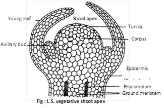
(iii) According to function : Haberlandt in 1890 classified the primary meristem at the apex of stem under the following three types :
(a) Protoderm : It is the outermost layer of the apical meristem which develops into the epidermis or epidermal tissue system.
(b) Procambium : It occurs inside the protoderm. Some of the cells of young growing region which by their elongation and differentiation give rise to primary vascular tissue, constitute the procambium.
(c) Ground meristem : It constitute the major part of the apical meristem develops ground tissues like hypodermis, cortex, endodermis, pericycle, pith and medullary rays.
(iv) According to plane of cell division : On the basis of their plane of cell division meristem are classified into three categories :
(a) Mass meristem : The cells divide anticlinally in all planes, so mass of cells is formed. e.g., formation of spores, cortex, pith, endosperm.
(b) Plate meristem : The cells divide anticlinally in two planes, so plate like area increased. e.g., formation of epidermis and lamina of leaves.
(c) Rib or File meristem : The cells divide anticlinally in one plane, so row or column of cells is formed. e.g,, formation of lateral root.
(2) Structure and organisation of apical meristem
(i) Vegetative shoot apex : Shoot apex was first recognized by Wolff (1759) shoot apex is derived from meristem present in plumule of embryo and occurs at the tip of stem and its branches as terminal bud. It also occurs in the inactive state in the axils of leaves as lateral buds. The tip of the shoot apex is dome-shaped and from its flanks at the base of the dome divide to form one or more leaf primordia. This continues throughout the vegetative phase. Many theories have been put forward to explain shoot apex, such as :
(a) Apical cell theory : This theory was proposed by Nageli (1858). According to this theory, shoot apical meristem consists of single apical cell. This theory is applicable in case of higher algae, bryophytes and in many pteridophytes but not in higher plants (i.e., gymnosperms and angiosperms).
(b) Histogen theory : It was proposed by Hanstein (1870). According to this theory, the shoot apical meristem consists of three distinct meristematic zones or layers (or histogens).
·
(c) Tunica corpus theory : This theory was proposed by Schmidt (1924). According to this theory, the shoot apex consists of two distinct zones.
(ii) Root apex : A group of initial cells, present at the subterminal region of the growing root tip, which is protected by a root cap is called root apical meristem or root apex. It is embryonic in origin and formed from the radicle part of embryo. However, in adventitious roots it is produced from derivatives of root apex. The root apex differs from shoot apex as it is short and more or less uniform due to complete absence of lateral appendages (leaves and branches) and differentiation of nodes and internodes. According to Hanstein (1870) root apex of most of the dicotyledons also consists of three meristematic zones - plerome, periblem and dermatogen (fourth meristem calyptrogen to form root cap only in monocots). Regarding the apical organisation of root following theories have been put forward.
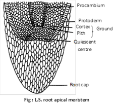
(a) Korper-Kappe theory : It was proposed by Schuepp (1917). This theory is comparable with the tunica and corpus theory of shoot apex. Korper means body and Kappe means cap.
(b) Quiescent centre theory : It was proposed by Clowes (1961). According to him, in addition to actively dividing cells, a zone of inactive cells is present in the central part of the root apex called quiscent centre.
The cells in this region have light cytoplasm, small nuclei, lower concentration of DNA, RNA and protein. These cells also contain fewer number of mitochondria, less endoplasmic reticulum and small dictyosomes.
Types of root apex : It is divided into following four types :
Common monocot root : Root apex is made up of four layers of histogen.
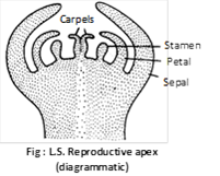
(iii) Reproductive apex : During reproductive phase, the vegetative apices are converted into reproductive apices. Before conversion, the apex stops producing leaf primordia. The summit of the apex which remained inactive during the vegetative phase, starts dividing. As a result of cell divisions, the apical meristem undergoes change in shape and increase in size. The apex may develop into a flower or an inflorescence. When the apex is to develop into a single flower, the cells at the flanks of the apex produce sepals and petals while the cells in the centre of summit produce stamens and carpels.
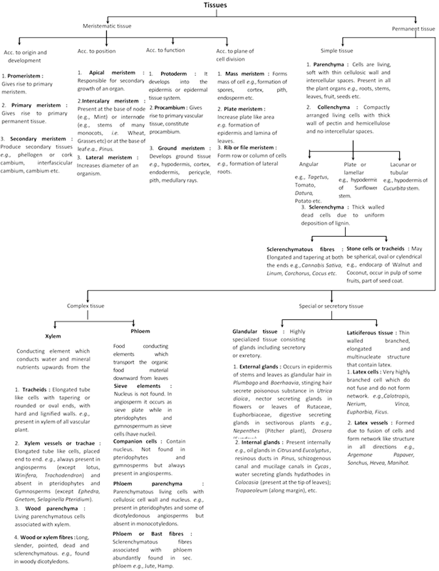
Permanent tissues
Permanent tissues are made up of mature cells which have lost the capacity to divide and have attained a permanent shape, size and function due to division and differentiation in meristematic tissues. The cells of these tissues are either living or dead, thin-walled or thick-walled. Permanent tissues are of three types :
(1) Simple tissues : Simple tissues are a group of cells which are all alike in origin, form and function. They are further grouped under three categories :
(i) Parenchyma : Parenchyma is most simple and unspecialized tissue which is concerned mainly with the vegetative activities of the plant.
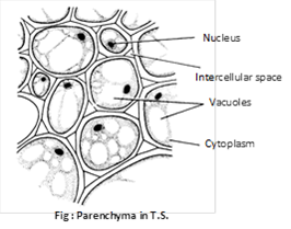
The main characteristics of parenchyma cells are:
(a) The cells are thin-walled and soft.
(b) The cells usually are living and possess a distinct nucleus.
(c) The cells contain well-developed intercellular spaces amongst them.
(d) The cytoplasm is vacuolated and cell wall is made up of cellulose.
(e) The shape may be oval, spherical, cylindrical, rectangular and stellate (star shaped) in leaf petioles of banana and canna and some hydrophytes.
(f) This tissue is generally present in almost all the organs of plants, i.e., roots, stems, leaves, flowers, fruits and seeds.
(g) If they enclose large air spaces they are called as aerenchyma; if they develop chlorophyll, they are called as chlorenchyma and if they are elongated cells with tapering ends, they are called as prosenchyma.
Functions: They perform the following functions:
(ii) Collenchyma : The term collenchyma was coined by Schleiden (1839). It is the tissue of primary body. The main characteristics of are given below :
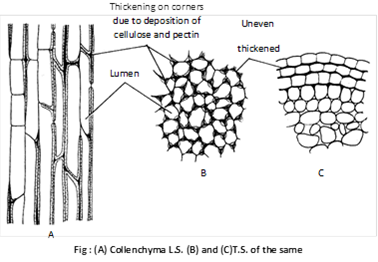
Collenchyma occurs chiefly in the hypodermis of dicotyledonous stems (herbaceous, climbers or plants e.g. Cucurbeta, Helianthus) and leaves. They are usually absent in monocots and in roots.
Special or Secretory tissues
These tissue perform special function in plants, e.g., secretion of resins gum, oil and latex.
These tissues are of two types :
(1) Laticiferous tissues
(2) Glandular tissues
(1) Laticiferous tissues : They are made up of thin walled, elongated, branched and multinucleate (coenocytic) structures that contain colourless, milky or yellow coloured juice called latex. These occur irregularly distributed in the mass of parenchymatous cells. latex is contained inside the laticiferous tissue which is of two types :
(i) Latex cells : A laticiferous cell is a very highly branched cell with long slender processes ramifying in all directions in the ground tissue of the organ. They do not fuse and do not form network. Plants having such tissues are called simple or non-articulated laticifers. e.g., Calotropis (Asclepiadaceae) Nerium, Vinca (Apocyanaceae), Euphorbia (Euphorbiaceae), Ficus (Moraceae).
(ii) Latex vessels : They are formed due to fusion of cells and form network like structure in all directions. At maturity, they form a highly ramifying system of channels full of latex inside the organ. Plants having such tissues are called compound or articulated laticifers. e.g., Argemone, Papaver (Papaveraceae), Sonchus (Compositae), Hevea, Manihot (Euphorbiaceae).
(2) Glandular tissue : This is a highly specialized tissue consisting of glands, discharging diverse functions, including secretory and excretory. Glands may be external or internal.
(i) External glands : They are generally occur on the epidermis of stem and leaves as glandular hair in Plumbago and Boerhaavia, stinging hair secrate poisonous substance in Urtica dioica, nectar secreting glands in flowers or leaves. e.g., Rutaceae and Euphorbiaceae. Digestive enzyme secreting glands in insectivorous plants e.g., Drosera (Sundew), Nepenthes (Pitcher plant).
(ii) Internal glands : These are present internally and are of several types. e.g., oil glands in Citrus and Eucalyptus, resinous ducts in Pinus, mucilage canals in Cycas. Water secreting glands (hydathodes) in Colocasia (present at the tip of leaves), Tropaeoleum (along margin), etc. The glands which secrete essential oil are called osmophores (osmotrophs).


You need to login to perform this action.
You will be redirected in
3 sec

