Category :
NEET
Gametogenesis
The process of the formation of haploid gametes from the undifferentiated, diploid germ cells in the gonads for sexual reproduction is called gametogenesis.
- As a result of this process, male gamete sperm and female gamete egg is formed.
- The process of Gametogenesis is stimulated by the FSH or Follicle Stimulating Hormone and for this process Vitamin "A" and "E" are also necessary.
- The process of Gametogenesis has been divided into 3 substages -
(1) Multiplication phase.
(2) Growth phase.
(3) Maturation phase.
Types of gametogenesis
(i) Spermatogenesis
- The process of formation of sperms in the germinal-epithelium of the testis of the male animal is termed as spermatogenesis.
- In mammals, testis have several coiled tubules in it called the seminiferous tubules.
- Sperms are formed in these tubules. The inner wall of seminiferous tubules is made up of germinal epithelium whose cells are cuboidal.
- Some special cells are present in this germinal epithelium which are called the primordial germ cells. Due to the division of these cells sperms are formed.
- Some large cells are also found in this germinal epithelium. These are called the "Sertoli cells or Subtentacular cells".
- These cells provide nutrition to the maturing sperms in the form of Glycogen.
- For getting nutrition, the head of the sperms are submerged in the cytoplasm of sertoli cells.
- When sperms fully mature, they move away from sertoli cells and get liberated in the cavity of seminiferous tubules.
- Liberation of sperms from sertoli cells is termed as Spermiation.
- Liberation of sperms from the testis is termed as Semination.
- Liberation of sperms into the vagina of the female is termed as Insemination.
- Sertoli cells are also endocrine in nature and they secrete 2 hormones -
AMH (Anti Mullarian Hormone): This hormone stimulates degradation of female gonads in a male embryo.
Inhibine hormone: This hormone is secreted in adult stages and it stops the secretion or FSH.
- Sertoli cells mainly provide nutrition and conserve the various stages of spermatogenesis. Spermatogenesis is a continuous process. To make it easier for study, it has been divided into the following steps -
(a) Formation of spermatid.
(b) Spermiogenesis or Spermatolesis.
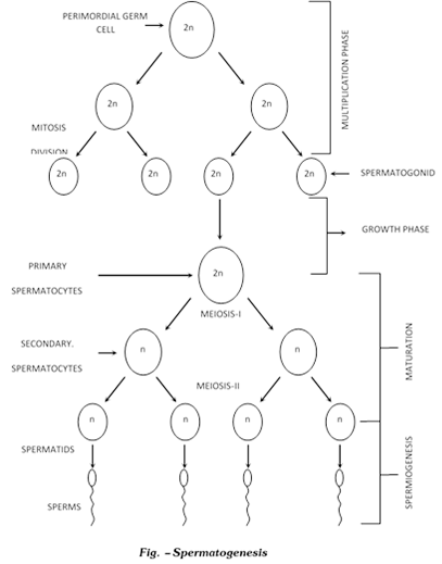
(a) Formation of spermatids: This process begins as the animal attains sexual maturity. The cells of the germinal epithelium of the seminiferous tubules which participate in this process are termed as the primordial germ cells. The process of formation of spermatids from primordial germ cells are termed as spermatocytosis. It has 3 sub-stages -
(1) Multiplication phase: During this process the primordial germ cells repeatedly undergo mitosis division, and as a result of these divisions spermatogonia are formed spermatogonia are diploid.
(2) Growth phase: Some spermatogonia either due to growth or due to food storage become 2 or 3 times of their original size, and are now known as primary spermatocytes. The remaining spermatogonia remain in the seminiferous tubules in the form of reserved stock. The primary - spermatocytes formed during the growth phase are diploid. Growth phase is the longest.
(3) Maturation phase: Primary - spermatocytes undergo Meiosis-I and as a result 2 haploid secondary spermatocytes are formed. This division is termed as First Maturation Division or Reductional division. Secondary spermatocytes undergo Meiosis II or equational division, and as result, 2 spermatids are formed from each secondary spermatocyte. Thus, from 1 diploid primary spermatocytes 2 secondary spermatocytes are formed on meiosis I and from 2 haploid secondary spermatocytes 4 spermatids are formed on meiosis-II. Metamorphosis of spermatids into sperms in known as Spermiogenesis or Spermatoliosis.
(b) Spermatoliosis: The process of transformation of a round non-motile and haploid spermatid obtained from spermatocytosis into thread-like, motile and haploid sperm is termed as spermatoliosis. From different parts of the spermatid different parts of the sperm are formed. These are as follows -
(i) From nucleus and glogibody -> Head part
(ii) From mitochondria -> Middle part
(iii) From distal centriole -> Tail part.
(i) The structure of the head of the sperm mainly depends on the structure of the nucleus. During spermatoliosis, nucleus contracts and acquires different shapes.
- RNA and nucleolus disappear from nucleus or their major part is given out from the nucleus. DNA also contracts / shrinks e. now the nucleus contains only those materials which are responsible for the hereditery characters.
- Centrosome divides into 2 centrioles e. 1 distal and 1 proximal centriole.
- Many golgi-bodies aggregate on the posterior side and their vacuoles enlarge.
- In some vacuoles of the golgi-bodies. Some dense-bodies can be seen, which are termed as Acroblast.
- In any one vacuole the Acroblast enlarges by aggregating with other. These are termed as “Acrosomal Granules”.
- The rest part of the golgi-body is given out and this is termed as Golgi-rest.
- This acroblast reaches the anterior most tip of the sperm and acquires a cap shaped structure on the nucleus which is termed as the Acrosome.
- Acrosome is surrounded by a double membrane i.e. the tonoplast and cell-membrane.
- The acrosome along with this membrane is termed as Galea-capatis. Acrosome, at the tip of the nucleus has an important role in breaking the egg.
- Mitochondria from different parts of spermatid, collect around the growing axonema and form a dense structure. In mammals, they form a spiral structure called Nebenkern sheath. In other animals, they collect and form mitochondrial clumps.
- The part of the axonema surrounded by mitochondria is known as Middle-piece.
- Around the middle-piece cytoplasm is present in the form of a thin layer called Manchette.
- Behind the middle piece, axonema surrounded only by plasmalemma is present. This is called the tail part.
- Shape of the sperm is so that it can move easily in a liquid medium.
- Structures and functions of sperms parts -
(a) Acrosome : Breaking the egg
(b) Middle piece : For providing energy
(c) Tail : For locomotion
Above structures are formed during spermatoliosis and the remaining substances are given out in the form of cytoplasmic residue, which is ingested by the "Cells of Sertoli".
(ii) Structure of sperm: Structure of sperm has three parts
(a) Head
(b) Middle piece
(c) Tail
(a) Head: It is flat ant oval in human sperm. It is composed of a large posterior nucleus and a small anterior acrosome.
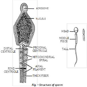
Acrosome
- Acrosome is formed from the golgi complex. It contains digestive enzyme "spermlysin". It is the caplike covering above the nucleus. It is surrounded by double membrane. Acrosome + its membrane are together called Galea-capatis. Acrosome plays important role in penetration of ovum by sperm.
- Remaining part of the head is nucleus. Narrow space between the nucleus and the acrosome is termed as "perforatorium".
- Nucleus of the sperm is very small. In it nucleoplasm and nucleolus are absent. It contains only chromatin.
- At the base of the nucleus in a pit like depression proximal centriole is present.
- In between the head and the middle piece a small neck is present. In this neck part a distal centriole is located. Both the centrioles are at right angles to each other.
- Proximal centriole first induce cleavage in a fertilized First spindle fibre forms from it.
- Distal centriole gives rise to the axial filament of the sperm. It has (9+2) microtubular arrangement.
(b) Middle piece
- This is known as the energy-chamber of the sperm. Many mitochondria spirally surround the axonema, this is called "Nabenkern sheath". This part provides energy to the sperm for locomotion.
- In middle-piece, cytoplasm is found in the form of a thin-sheet called Manchett.
- In middle-part, axonema is surrounded by 9 solid fibres made up of proteins.
- At the posterior end of the middle-piece a Ring centriole is found. Its function is not known.
(c) Tail
- The longest and the fibrous part of the sperm is termed its tail.
- Sperm moves with the help of its tail.
- Basal granule of the tail is Distal centriole.
Tail has 2 parts
- Main part : This part is broad. It contains cytoplasm and is surrounded by 2 solid fibres.
- End piece : This part is narrow in it cytoplasm is absent only axonema is present. In it solid fibres are also absent. In the sperm of certain animals, tail is absent. g.
(1) Ascaris : Tailless, ameboid sperms
(2) Cray fish : Tailless, stellate (star shape) sperms.
(3) Crab and lobser : Tailless sperms with 3 spines at apex.
(4) Biflagellage sperms : In Toad fist (Opsanus)
(5) In Opposum : Many sperms fuse together by their heads to form a "sperm-boat".
(6) Gastrapods have hexaflagellated sperms.
(iii) Oogenesis : Oogenesis takes place in the ovaries. Unlike sperm formation that starts at puberty, egg formation begins before birth but is completed only after fertilization. Oogenesis consists of three phases -
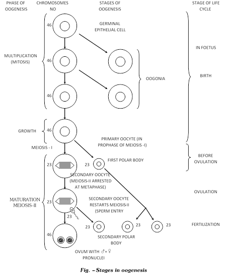
- Multiplication phase
- During foetal development, certain cells in the germinal epithelium of the ovary are larger than others and also have larger nuclei.
- These cells undergo mitotic divisions, producing undifferentiated germ cells called oogonia or egg mother cells in the ovary.
- The oogonia have diploid, number of chromosome, 46 in humans.
- The oogonia multiply by mitotic divisions and produce ovigerous cords or egg tubes of pfluger in mammals.
(b) Growth phase: It is prolonged and slow. Oogonia form rounded masses or egg nests at the tips of egg tubes of pfluger.
- An egg nest forms ovarian follicle (Graffian follicle) one central oogonium grows and functions as primary oocyte. The others form the covering follicular cells. the latter provide nourishment to primary oocyte. Some nourishment also comes from outside. Yolk is deposited in this state. This phenomenon is called vetellogenesis.
- In cooperation with follicular cells, the enlarged primary oocyte secrete mucoprotein membrane or zona pellucida outside its own plasma membrane or vitelline membrane. There is increase in reserve food, size of nucleus, number of mitochondria; functioning of golgi apparatus and complexing of endoplasmic reticulam.
(c) Maturation phase
- Meosis occurs. Nucleus shifts towards animal pole and undergoes meosis - I. A daughter nucleus alongwith small quantity of cytoplasm is extruded as primary polar body or polocyte below zona pellucida. Simultaneously primary oocyte is changed into haploid secondary oocyte. It proceeds with meosis - II but stops at metaphase-II. Ovum is generally shed in secondary oocyte stage.
- After fertilization, the second meotic division is completed with unequal cytoplasmic cleavage. This forms a large cell the ootid with essentially whole of the cytoplasm, and a very small cell, the second polar body. The ootid and the second polar body are haploid as the second meotic division is equational. The first polar body may divide at about the same time into two polar bodies.
- One primary oocyte forms, after two meiotic division, one haploid ootid and three haploid polar bodies. The ootid grows into a functional haploid ovum.
- The polar bodies have no function and disintegrate due to lack of cytoplasm and food.
- The formation of non functional polar bodies enables the egg to get rid of excess chromosomes. The unequal cytoplasmic division enables the ovum to retain the whole of cytoplasm of the primary oocyte in it for the development of the future embryo.
- In humans, ova are released from the ovary in the secondary oocyte stage. Their maturation is completed in the mother's genital tract, usually after the sperm has entered for fertilization.
(iv) Structure of ovum: An ovum is generally spherical, nonmotile gamete with yolky cytoplasm and enclosed in one or more egg envelops. Size of ovum varies in different animals and depends upon the amount of yolk. Size of ovum varies from 10m to a few cm. Largest sized egg is of ostrich and is about 170 ´135 mm. Egg size and yolk amount are interdependent. It is about 50m in many polychaete worms, 150m in tunicates but very large sized in birds and reptiles. In mammals, it is generally microlecithal and about 100m.
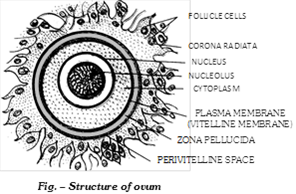
Human ovum is microlecithal with large amount of cytoplasm. Cytoplasm is differentiated into outer, smaller and transparent exoplasm or egg cortex and inner, larger and opaque endoplasm or ooplasm.
Egg cortex is with some cytoskeletal structures like microtubules and microfilaments (Balinsky, 1981), pigment granules and cortical granules of mucopolysaccharides. Endoplasm is with cell-organelles, informosomes tRNAs, histones, enzymes etc. Nucleus of ovum is large, bloated with nucleoplasm and is called germinal vesicle. Nucleus is excentric in position so human ovum has a polarity. The side of ovum with nucleus and polar body is called animal pole, while the opposite side is called vegetal pole.
Egg envelopes. Human ovum is surrounded by a number of egg envelopes:
(a) Vitelline membrane: It is inner, thin, transparent and is secreted by ovum itself.
(b) Zona pellucida: It is middle, thick, transparent and non-cellular.
(c) Corona radiata: It is outer, thicker coat formed of radially elongated follicular cells. Between the vitelline membrane and zona pellucida, there is a narrow perivitelline space.
Differences between Spermatogenesis and Oogenesis
| S. No. |
Characters |
Spermatogenesis |
Oogenesis |
|
1.
|
Site of occurrence
|
In the seminiferous tubules of testes.
|
In the ovaries.
|
|
2.
|
Total period
|
It is a continuous process and completed in 74 days in humans
|
It is a discontinuous process and completed in a few days to years.
|
|
3.
|
Growth phase
|
Of shorter duration
|
Of longer duration
|
|
4.
|
Yolk synthesis
|
No yolk is synthesized in growth phase
|
Vitellogenesis occurs in growth phase.
|
|
5.
|
Nuclear changes
|
Nucleus becomes condensed by the loss of superfluous materials.
|
Nucleus is bloated due to increase in necleoplasm.
|
|
6.
|
Number of gametes
|
One spermatogonium forms 4 haploid sperms.
|
One oogonium forms only one ovum.
|
|
7.
|
Polar bodies
|
Not formed.
|
Two or three polar bodies are formed.
|
|
8.
|
Site of completion
|
It is started and completed within the testes.
|
It is started inside the ovary but is generally completed outside the ovary.
|
|
9.
|
Size of gametes formed
|
Sperm is much smaller than spermatogonium.
|
Ovum is much larger than oogonium.
|
(vi) Types of eggs
(a) On the basis of amount and distribution of yolk
(1) Alecithal or Microleithal or Oligolecithal or Meolecithal and Isolecithal or Homolecithal: The amount of yolk is very small in these types of eggs. (Oligolecithal or Microlecithal or Alecithal) and yolk is evenly distributed in these eggs (Isolecithal or Homolecithal). Examples - Egg of Amphioxus, Eutheria (Human egg), Metatheria and Sea-urchin.
(2) Mesolecithal or Telolecithal eggs: In this type of egg the amount of yolk is moderate and yolk is concentrated in the basal part of egg (telolecithal egg). Examples - Egg of Amphibia, Petromyzon and Lung fishes.
(3) Polylecithal or Macrolecithal or Megalecithal eggs: Eggs are with large amount of yolk and this yolk is concentrated in the centre and cytoplasm is in the form of superficial layer around the yolk (centrolecithal eggs). Example - Insect's egg.
In discoidal or highly telolecithal eggs, the yolk is enormous in amount and cytoplasm is confined to a disc like area on yolk. This disc of cytoplasm is called germinal disc. Example - Eggs of reptiles, birds, protherian mammals.
Note: Majority eggs are oval but the eggs of insects are long and cylindrical.
- Smallest eggs are of 50m in the polychaeta and the largest eggs are of an ostrich.
(b) On the basis of fate
(1) Determinate / Mosaic eggs: Every part of fertilize egg has a definite fate, so that fate of every blastomere is determined from the beginning. It is found in invertebrates except echinoderms.
(2) Indeterminate / Regulative eggs: The fate of different parts of egg or its blastmeres is not predetermined. Example - Echinodermes, Vertebrates.
(c) On the basis of shell
(1) Cleidoic eggs: Eggs surrounded by a hard shell are know as cleidoic eggs. These eggs are found in those animals which have a terrestrial mode of life of which lay eggs on land. These eggs have more amount of yolk. These are adaptations to terrestrial mode of life. Shell prevents the egg from dessication. e.g. - Eggs of "Reptiles". "Birds". "Insects" and "Prototherians".
(2) Non - Cleidoic eggs: Eggs which are not surrounded by a hard shell are called Non-cleidoic eggs. These eggs are found in all oviperous animals which lay eggs in water and all viviperous animals. e.g. - All viviperous animals (Mammals) and all oviperous animals which lay eggs in water (Amphibians).
Structure of an oocyte: Oocyte is a large yolk containing cell. The nucleus is large and swollen and is termed as the "Germinal Vasicle". Oocyte is surrounded by membranes termed as the egg-membranes. Oocyte / Ovum alongwith the egg-membrane are termed as the egg. Egg = Ovum / Oocyte + Egg membrane.
Classification of egg - membranes: On the basis of origin, egg-membranes are of 3 types -
(1) Primary egg membrane: This membrane is secreted by the egg (ovum) itself. e.g. - Vitelline membrane, of human egg.
(2) Secondary egg membrane: This is found outside the primary egg membrane and is secreted by the ovary. e.g. Chorion of insect eggs, corona radiata and zona pellucida of human egg.
(3) Tertiary egg membrane: This present outside the primary and the secondary egg membrane. It is either secreted by the uterus or the oviduct. Egg jelly coat around frog's egg; albumen, shell membrane and shell of bird egg.
Functions of egg membranes
(1) To provide protection.
(2) To check polyspermy.
(3) To provide buoyancy to the amphinian eggs.
(4) To provide nutrition (Birds, Reptiles)
(5) To help in excretion (Allantois)
Different types of eggs
(1) Insect egg: Eggs of insects are megalecithal or polylecithal in them yolk is present in the centre, so the eggs are also centrolecithal. Eggs of insects are cigar like. Along with plasma-membrane the egg has 2 egg-membranes.
(i) Vitelline membrane: This is a primary egg membrane and the egg itself secretes it around.
(ii) Chorion: This is a secondary egg membrane and is secreted by the ovary. In Insect's egg tertiary egg-membranes is absent. Chorion of insect's egg is ornamented i.e. there are specific markings on its egg membrane which are characters of Taxonomic importance. In the egg, a hole termed as micro Pyle is present which is the port of entry for sperms. Its cytoplasm is divided into 2 parts -
(a) Central
(b) Peripheral cytoplasm

(a) Central cytoplasm: It is present in a very small amount in the centre of the egg. Egg nucleus is located in it.
(b) Peripheral cytoplasm: It is present in a very small amount along the periphery of the egg.
Yolk: In insect's egg yolk is present in a very large amount and this yolk is concentrated between the central and the Peripheral cytoplasm.
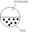
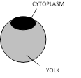
(2) Frog's egg: Eggs of frog are Teloecithal and Mesolecithal. The egg has 2 egg membranes.
(i) Vitelline membrane: This is a primary membrane, secreted around by the egg itself.
(ii) Jelly - coat: This is a tertiary egg-membrane. It is secreted by the oviduct. Secondary egg-membrane are absent in these egg's. Internally, the egg is divided into 2 areas -
(a) Animal pole (b) Vegetal pole
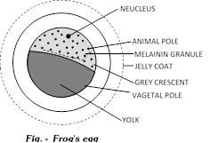
(a) Animal pole: This part has more amount of cytoplasm in it and the egg nucleus is also located in it. In this part melanin granules are found which prevent the egg from harmful radiations. Due to these melanin granules the frog's egg is partly white and partly black. This helps in Camouflage. Sperm always enters inside the egg through the animal pole. The part from where the sperm enters inside the frog's egg in future forms the ventral part of the embryo. As the sperm enters inside the egg. The part directly opposite to the entry point becomes a clear-zone due to the rapid movement of melanin granules this clear-zone is termed as the Grey-Crescent. This part with Grey-Crescent forms the dorsal part of the embryo in future.
(b) Vegetal pole: Here the yolk is concentrated in frog's egg, the part with cytoplasm in future forms the ectoderm. The Grey crescent part in future the Mesoderm and the part with yolk in future forms the endoderm.
Jelly-coats of all the eggs of a frog absorb water and swell up, to form a cluster of eggs termed as Spawn. Jelly-coat has air-bubbles, due to which the eggs don't drown. Jelly-coat is bilter in taste and so the eggs are protected from the enemies.
Important tips
- The longest phase of gametogenesis is growth-phase.
- Acrosome of the sperm is formed by the golgi-body.
- Smallest sperm is of Crocodile (.02mm) and largest sperm is of discoglossus (2mm)
- Complete spermatogenesis in man takes place in 74 days.
- In 1ml of human semen 100 million sperms are present.
- Infertility which arises die to less number of sperms is called Oligospermia.
- Condition in which sperms are totally absent in semen is also termed as Azospermia.
- In oogenesis, yolk is synthesised in the growth phase.
- Largest egg is of Ostrich which along with the shell is of 16 cms.
- Length of a human sperms in approx 55me.
- The condition of presence of normal number of motile sperms in human semen is termed as isozospermia.
- The condition of presence of completely non-motile sperms in human semen is termed as Necrospermia.
- Smallest eggs are of humming bird.
- In lower animals large amount of eggs are poduced because their chances of survival are very less.
- Order of egg-production. Mammals < Birds < Repitles < Amphibia < Pisces.
- Cat and Rabbit are both induced ovulators.
- The life span of eggs in female reproductive organs is different e.g. in humans it is 48 hours.
- Nucleus of the egg is termed as Germinal vesicle.
- The asexual process replaced by the sexual method is known as apomixis.
- Phallic organs in cockroach are related to male reproductive system.
- No natural death in organisms showing binary fision e.g. Amoeba, so are called immortal.
- The croaking sound made by frog is sex call for female partner.
- Leuvenhock (1677) saw human sperm.
- In frog bidder canal help in sperm passout.
- Gynandromorph : An animal having male characteristics on one part and female characteristics on the
other.
- Gynaecomastia: Enlarged functional mammary gland in male.
- Azoospermia: No sperms in semen.
- Oligospermia : Sperms less than 20 million in per ml semen.
- V. Graf (1672): Androgenesis discovered follicles in human ovary.
- Androgenesis: Development in which embryo has only paternal chromosomes, male parthenogenesis.
- Gynogenesis: Development in which embryo has only maternal chromosomes, female parthenogenesis.
- Gland of Tyson: Modified sebaceous glands present around corona of glans.
- In many birds (exception some birds of prey) only the left ovary and left oviduct are function. The right ones are non-functionsl.
- Seminiferous tubules: Structural and functional unit of testes.
- Hysterectomy: Surigcal removal of uterus.
- Castration / Chidectomy: Removal of testes. It produce eunuchs. Castration changes aggressiveness of male into docile nature.
- Corpus luteum: Persists for two weeks in case of non-pregnancy and four months when pregnancy has taken place.
- Prostatitis: Inflammation of prostate gland. Prostate cancer is common in ageing males.
- Human egg:1 mm in diameter.
- Prostatectomy: Surgical removal of prostate gland.
- Peculiar spermatozoa: Ascaris has amoeboid spermatozoa devoid of flagellum. Some crustaceans also have atypical sperms.
- Sperms form about 10% of the ejaculated semen.
- Protandry: Spermatozoa mature earlier than ova in bisexual animals e.g. - Hydra, Earthworm.
- Andrology: Branch of medicine concerned with diseases peculiar to male sex.
- Spermatorrhoea: An involuntary discharge of semen, without orgasm.
- Spermatophore: A capsule containing spermatozoa, as in cuttle fish and salamander.
- Menarche: Beginning of menstrual cycle and other bodily changes.
- Oophoritis: Inflammation of an ovary.
- Vitellogenesis: Process of laying down of yolk in the primary oocyte. It occurs in the prophase of meosis-I.
- Metagenesis: Alternation of sexually and asexually reproducing forms in the life cycle of an animal e.g. Obelia.
- Protogyny: Ova mature earlier than sperm in abisexual animal e.g. Herdmania.
- Spermathecae: Small sacs that form a part of female reproductive system of earthworm and store spermatozoa received from the male for use in future.
- Oviparous: Animals that lay eggs e.g. - Birds.
- Viviparous: Animals which give birth to young ones, e.g. most mammals.
- Ovoviviparous: Animals that produce eggs which hatch within their bodies.
- Ovipositor: A specialised female organ for laying eggs, specially in insects.
- Rutting season: It is a brief period of pronounced sexual activity in males.
- Tubectomy (Salpingectomy): Surgical removal of oviducts.
- Von bear: Discovered ovum.
- Strobilation: Asexual multiplication by transverse fusion and is found in Scyphistoma of Aurelia and also found in Taenia.
- Richard owen gave term parthinogenesis.
- Testes are also called spermaries.
- Vaginal coelom: Cavity of scrotal sac.
- Leydig cells are absent from the testes of frog and are characteristics of mammalian testes.
- To-gene: Testicular organisation gene located on Y-chromosome and is a male determining factor.
- Adiposogenital syndrome: Hypogonadism in male and characterised by obesity and child like sexual organs.
- No man - no woman syndrome: Characterised by male-female pseudohermaphroditism in which external sexual characters are opposite to genetic and gonadal sex.
- Wolffian duct: Acts as male genital duct, while mullarian duct is vestigeal.
- Mesosalpinx: Mesentry suspending the fallopian tube.
- Mesometrium: Mesentry suspending the uterus.
- Uterus: It is also called womb.
- Cervix of uterus is formed of most powerful smooth muscles.
- Vestibule: Acts as a urinogenital sinus.
- Perineum: Area between the fourchette and anus.
- Bartholin's or Bulvo vestibular glands of female homolegous to Cowper's glands of male.
- Precocious puberty: Puberty attained before the normal age.
- Hypermastia: More than normal number of breasts.
- Number of breasts in female depends upon the number of young ones born at a time.
- Female ascaris has paired ovaries so is called didelphic.
- In seasonally breedings animals, testes show testicular cycle.
- Spermatogenesis is continous process, while oogenesis is a discontinuous process.
- In spermatogenesis, spermatogonium produces four sperms while in oogenesis, one oogonium produces one ovum and 2 or 3 polar bodies.
- Golgi rest: Part of golgi body which is lost during spermiogenesis.
- Gynosperm: Sperm with 22 A+X chromosome.
- Androsperm: Sperm with 22 A+Y chromosome.
- Yolk nucleus: Also called Balbiani body. A mass of mitochondria and golgibody near the nucleus which controls vitellogenesis.
- Redundancy: Gene amplification of r-RNA genes for rapid RNA and protein synthesis.
- Ring centriole: Also called annulus or Jensen's ring. Menstruation is also called "Weeping of uterus for the lost ovum or funeral of unfertilized egg".
- Menstrual cycle is associated with withdrawal or progesterone.


