Current Affairs 11th Class

Vegetative reproduction (1) By budding : According to Bisset and Hale, reproduction by budding takes place in Bigidi bacterium bifidus. (2) By binary fission : This type of reproduction is most common in all kinds of bacteria. Under favourable conditions bacterial cell expands. Cytoplasm divides into two parts due to constriction and formation of a transverse septum in the centre of the cell. Later on, these two parts separate from each other and give rise to two cells. The speed of binary fission is decreased due to low temperature. Therefore, food is preserved in the cold storage. The cause of food spoilage and bacterial infection is the rapid multiplication of bacteria. Asexual reproduction (1) By endospores : Endospores are formed in all species of the genera Bacillus and Clostridium. In each cell only one endospore is formed. Endospore is highly resistant to very high and very low temperature. Endospore is found either in the centre or near the cell wall. Under unfavourable conditions cytoplasm shrinks and gets rounded and around it a hard protective three layer is formed. Each endospore may be either circular, ellipsoidal or semicircular. When favourable conditions come, outer layers rupture and active bacterial cell comes out. So this is a method of perennation (i.e., to tide over unfavourable condition) and some people say it “reproduction wihtout multiplication”. (2) By conidia : Some filamentous bacteria e.g., Streptomyces  reproduce by means of conidia. The conidia are spore like in structure and are formed in chains. Each conidium gives rise to a new bacterium. (3) By zoospores : In rare cases bacterial cell forms some motile spores which give rise to new cells. This process has been rarely seen. e.g., Rhizobium. Sexual reproduction (Genetic recombination) Sometimes it was believed that sexual reproduction does not take place in bacteria. Lederberg and Tatum (1946) proved that sexual reproduction takes place in bacteria. On the basis of this discovery they were awarded Nobel Prize. According to the present view, three types of sexual reproduction are found in bacteria : (1) Transformation : In this process one kind of bacterium is transformed into another kind. It takes place by transferring DNA from capsulated to non-capsulated bacterium. For the first time Griffith (1928) reported transformation in mice. Later on, Avery, Mcleod and McCarty (1944) studied transformation in Diplococcus pneumoniae. (2) Transduction : In this process DNA of a bacterial cell is transferred into another bacterial cell through bacteriophage – a kind of virus which is parasitic upon bacteria. Bacteriophage consists of DNA. It has been now accepted that DNA of a bacterial cell is transferred through bacteriophage to another bacterium. Transduction was first of all reported by N.D. Zinder and Lederberg (1952) in bacteria Salmonella typhimurium. (3) Conjugation : In this process genetic material from one strain of bacterium which is known as male is transferred into another strain of bacterium which is known as female. On the experimental basis it is believed that genetic material of male enters into female bacterium in the more...

With respect to oxygen requirement and mode of cellular respiration, bacteria distinctly belong to two broad categories : (1) Aerobic respiration Obligate aerobes : These bacteria grow exclusively in presence of molecular oxygen and fail to survive in its absence, e.g., Bacillus subtilis, Azotobactor, Arthrobactor, Mycobacterium etc. Facultative anaerobes : The aerobic bacteria which can also survive in absence of oxygen, e.g., Aerobacter, Klebsiella, Pseudomonas, etc. (2) Anaerobic respiration Obligate anaerobes : These bacteria grow and multiply in the absence of free oxygen. They fail to survive under aerobic conditions, e.g., Clostridium botulinum. Facultative aerobes : The anaerobic bacteria which can also survive in presence of oxygen, e.g., Chlorobium limicola.

They are gram negative obligate pleomorphic but walled obligate intracellular parasites which are transmissible from arthropods. They are intermediate between true bacteria and viruses. Rickettsias require exogenous factors for growth. Cell wall is like typical bacterial wall. ATP synthesis is absent but ADP is exchanged with host cell ATP. They have genome and size (0.3-0.5\[\mu m\]) smaller than true bacteria but have a longer generation time. Internally the cells of rickettsias contain DNA as well as RNA in a ratio of 1 : 3 .5. The cell walls contain muramic acid and are sensitive to lysozyme. Flagella, pili and capsule are absent. Reproduction occurs by binary fission. The natural habitat of rickettsiae is in the cells of arthropod gut. They cause typhus group of fevers. Spread by droplet method, lice, ticks, fleas, etc. Human diseases
Disease Casual organism
Typhus fever Rickettsia prowazekii.
Rocky mountain spotted fever Rickettsia rickettsii
Q fever Coxiella burnetti

These are free inhabitants of mud and water, and are chemoheterotrophic unicellular bacteria. These are spiral or helicoid in shape, covered by flexible cell wall. In spirochaetes flagella are absent but the cells are able to swim over solid surface by the fibrillae. Many diseases are caused by them as Treponema pallidum causes syphilis, Leptospira causes infectious jaundice and Borrelia causes relapsing fever. Besides some spirochaetes are found in teeth.

(1) Simple staining : The coloration of bacteria by applying a single solution of stain to a fixed smear is termed simple staining. The cells usually stain uniformly. (2) Gram staining : This technique was introduced by Hans Christian Gram in 1884. It is a specific technique which is used to classify bacteria into two groups Gram +ve and Gram –ve. The bacteria are stained with weakly alkaline solution of crystal violet. The stained slide of bacteria is then treated with 0.5 percent iodine solution. This is followed by washing with water or acetone or 95% ethyl alcohol. The bacteria which retain the purple stain are called as Gram +ve. Those which become decolourised are called as Gram –ve. In general the wall of Gram +ve bacteria have simpler nature as compared to Gram –ve bacteria. E.coli is a Gram –ve bacterium. Gram negative bacterium can be seen with other stain safranin.     Gram positive bacteria : e.g., Pneumococcus, Streptococcus, Staphylococcus, Bacillus, Clostridium, Mycobacterium, Streptomyces. Gram negative bacteria : e.g., Salmonella, Pseudomonas, Escherichia, Haemophilus, Helicobacter, Vibrio, Rhizobium.   
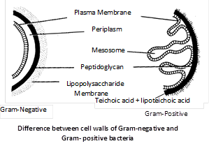
Gram -Positive Gram - Negative
Cell wall thick (250 - 300 Å). Cell wall thin (100 - 150 Å)
Cell wall homogenous. Cell wall heterogenous.
Cell wall single layered. Cell wall 3-layered.
Cell wall more rigid. more...
(1) Capsule : In a large number of bacteria, a slimy capsule is present outside the cell wall. It is composed of polysaccharides and the nitrogenous substances (amino acids) are also present in addition. This slime layer becomes thick, called capsule. The bacteria, which form a capsule, are called capsulated or virulent bacteria. The capsule is usually found in parasitic forms e.g., Bacillus anthracis, Diplococcus pneumoniae, Mycobacterium tuberculosis. It provides protection against phagocytosis and antibiotics. Capsule also protects the cell against dessication and viral attack. The capsulated bacteria are usually non-flagellated (i.e., Atrichous). Capsulated bacteria form smooth colonies and are known as S-type bacteria, which are highly virulent. Non-capsulated bacteria form rough colonies and are known as R–type bacteria. (2) Cell wall : All bacterial cells are covered by a strong, rigid cell wall. Therefore, they are classified under plants. Inner to the capsule cell wall is present. It is made up of polysaccharides, proteins and lipids. In the cell wall of bacteria there are two important sugar derivatives are found i.e., NAG and NAM (N-acetyl glucosamine and N-acetyl muramic acid) and besides L or D - alanine, D-glutamic acid and diaminopimelic acid are also found. One of the unique components of cell wall of bacteria is peptidoglycan or mucopeptide or murien (made of mucopolysaccharide + polypeptide). In peptidoglycan, NAG and NAM are joined by short peptide chains or cross bridges of amino acids. Outer layer of cell wall of Gram –ve bacteria is made up of lipopolysaccharides and cell wall of Gram +ve bacteria of teichoic acid. The cell wall of Gram positive bacteria is much thicker and contains less lipids as compared to that of Gram –ve bacteria. The enzyme lysozyme can dissolve the bacterial cell wall.     
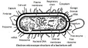
(3) Plasma membrane : Each bacterial cell has plasma membrane situated just internal to the cell wall. It is a thin, elastic and differentially or selectively permeable membrane. It is composed of large amounts of phospholipids, proteins and some amounts of polysaccharides but lacks sterols. It is characterised by possessing respiratory enzymes. Mesosome : On the plasma membrane generally at mid point, there are present some circular coiled bodies called mesosomes. So mesosomes are simply infoldings of plasma membrane. Mesosomes contain respiratory enzymes like oxidases and dehydrogenases and hence they help in respiration. Hence mesosomes are also known as "mitochondria of bacterial cell" or chondrioides. Mesosomes are more prominent in Gram +ve bacteria.
  • It receive DNA during conjugation and DNA replication enzyme.
  • Mesosome participate in the formation of septa during cell division.
(4) Cytoplasm and Cytoplasmic inclusions : The cytoplasm is a complex aqueous fluid or semifluid ground substance (matrix) consisting of carbohydrates, soluble proteins, enzymes, co-enzymes, vitamins, lipids, mineral salts and nucleic acids. The organic matter is in the colloidal state. The cytoplasm is granular due to presence of a large number of ribosomes. Ribosomes in bacteria are found in more...

Protista (Protistos = Primary) includes unicellular eukaryotes and show the following characters : (1) Protists include solitary unicellular or colonial unicellular eukaryotic organisms which not form tissues. (2) The unicells may be naked or covered by cell wall, pellicle, cuticle or shell. (3) Simple multinucleate organisms or stages of life cycles occur in a number of groups. (4) The organisms possess double and porous nuclear membranes, mitochondria, golgibody, plastids (in many), vacuoles, lysosomes and ribosomes is also present. Centrosome is occur in many cases. (5) In many forms, plastids, (9+2 strand) flagella and other organelles are present. (6) Some protists possess contractile vacuole for regulation of their water content. (7) Their reproductive cycles typically include both asexual divisions of haploid forms and true sexual processes with karyogamy and meiosis. (8) The organisms move by flagella or by other means or are non-motile. (9) It may be photosynthetic, holotrophic, saprotrophic, parasitic and symbionts. Some have mixotrophic nutrition (holotrophic + saprobic). The photosynthetic, floating protists are collectively called phytoplankton. The free-floating, holozoic protozoans are collectively termed zooplankton. (10) Asexual reproduction is the most common method in protists. It involve binary fission (Paramecium, Euglena, Amoeba), multiple fission (Amoeba), plastotomy (Opalina), budding (Paracineta, Arcella) and spore formation (Slime moulds). (11) Sexual reproduction is believed to have originated in primitive protists. It involve isogamy (Monocystis), anisogamy (e.g., Ceratium) and oogamy (e.g., Plasmodium). (12) Unicellular protists have been broadly divided in to three major groups : Photosynthetic protists : e.g., Dinoflagellates, Diatoms, Euglenoids. Consumer protists : e.g., Slime moulds or Myxomycetes. Protozoan protists : e.g., Zooflagellata, Sarcodina, Sporozoa, Ciliata.

Slime moulds include very interesting and peculiar organisms which share the characters of both animals and fungi. Due to this peculiarity they are commonly called fungus animals. Modern biologist include slime moulds under the kingdom-protista and called them protistan fungi. General characters (1) They do not have chlorophyll. (2) They are surrounded by the plasma membrane only (somatic parts are without cell walls). (3) At one stage of the life cycle they have amoeboid structure. (4) The slime moulds live usually amongst decaying vegetation. They are quite common on lawns and moist fields. (5) They exhibit wide range of colouration. (6) They have phagotrophic or saprotrophic nutrition. Parasitic forms are not known (7) The sporangia produce spores. Each spores possesses a cellulose cell wall. (8) The slime moulds resemble both protozoa and the true fungi. They are like protozoa in their amoeboid plasmodial stage and similar to true fungi in spore formation. Slime moulds are of two types : Acellular (Plasmodial) Slime moulds General characters (1) Acellular slime moulds commonly grow as slimy masses on damp places rich in dead and decaying organic matter. (2) The somatic phase is diploid and consists of a free living organic matter multinucleated protoplasm called plasmodium. (3) The plasmodium slowly streams or glides over decaying organic matter putting out blunt finger like pseudopodia showing amoeboid movement. (4) They also absorb dissolved organic substances from the substratum showing saprotrophic nutrition. (5) Under unfavourable conditions the plasmodium contracts and gets surrounded by thick horny wall. It is called sclerotium. (6) Each plasmodium reproduces asexually by the formation of several, small, sessile or stalked, brightly coloured sporangia. (7) The multinucleated protoplasm of sporangium is cleaved to produce a large number of small uninucleate spores. (8) When fully mature, the wall of the sporangium bursts to release the spores. The spores are dispersed by air. (9) On germination, a spore generally releases one biflagellate, spindle-shaped swarm cell or a non-flagellate myxamoeba. The myxamoebae feed on bacteria and yeasts and multiply in number. The swarm cells swim about actively and finally fuse in pairs at the posterior nonflagellate ends to form zygotes. (10) The diploid nucleus of zygote undergoes repeated mitotic divisions. As a result, the zygote gradually changes into a multinucleate amoeboid structure, the plasmodium. The plasmodium repeats the life cycle. Examples : Physarum, Physarella, Fuligo, Dictydium, Lucogala, Tubifera.      Cellular Slime moulds General characters (1) The cellular slime moulds occurs in the form of haploid uninucleated, naked (without cell wall) cell covered by plasma membrane. These cells are called myxamoebae. (2) The myxamoebae move freely with the help of amoeboid movement and phagotrophic or holozoic nutrition. (3) They grow and divide to form a large population of individuals. (4) Under unfavourable condition a myxamoeba secrete a rigid cellulose wall to form the microcyst. Microcyst formation is a means more...

  General characters (1) This is well defined group of unicellular, golden-brown photosynthetic organisms. Majority of them are motile and flagellated but a few are non-motile and non-flagellated. Flagellated forms exhibit peculiar spinning movement. Hence, they are called whorling whips. (2) The cell wall of dinoflagellates, if present, is composed of a number of plates made up of cellulose. It is called theca or lorica. The theca contains two grooves-longitudinal sulcus and transverse girdle or annulus. (3) Usually the cells possess two flagella which are of different types (heterokont). (4) Trichocysts are rod like or spindle shaped ejective structures which occur just below the cell membrane. (5) Cells possess a relatively large and prominent nucleus known as mesokaryon. DNA is without association with histone. (6) There are numerous discoid chloroplasts without pyrenoids. They are yellow-brown to dark-brown in colour due to presence of characteristic pigments – Chlorophyll a, c, \[\alpha \]- carotene and xanthophylls (including dinoxanthin and peridinin). (7) The reserve food material is starch in fresh water forms and oil in marine forms. (8) The cells possess an osmoregulatory organelle called pusule which superficially looks like contractile vacuole. The cells posses mitochondria, ribosomes and golgi bodies. They also possess mucilage bodies or vesicles below the cell membrane. (9) In dinoflagellates it is mainly holophytic or photosynthetic. However, some forms are saprobic, parasitic, symbiotic or holozoic. For example, an colourless Blastodinium is parasite on animals. (10) Dinoflagellates reproduce asexually through cell division or by the formation of zoospores and cysts. The cell division starts from posterior end. During cell division, centromeres and spindle are not seen. The spindle is replaced by cytoplasmic microtubules. During mitosis, the chromosomes break up into pairs of chromatids. The nuclear envelops and nucleolus persists during division. (11) If sexual reproduction occurs, is isogamous or anisogamous. Two cells conjugate by a conjugation canal where the two amoeboid gametes fuse to form a diploid zygote. Life cycle involves zygotic meiosis (e.g., Ceratium, Gymnodinium etc.) or gametic meiosis (e.g., Noctiluca). Examples : Glenodinium, Peridinium, Gymnodinium, Gonyaulax, Ceratium, Noctiluca.       General character (1) Most of the diatoms occur as phytoplanktons both in fresh and marine waters. A few forms occur as benthos the bottom of water reservoirs. Diatoms constitute a major part of phytoplankton of the oceans. (2) The cells of diatoms are called frustules or shell. They are microscopic, unicellular, photosynthetic organisms of various colours and diverse forms. They may be circular, rectangular, triangular, elongated, spindle-shaped, half-moon shaped, boat-shaped or filamentous. Incipient filament occur in Melosira. (3) They exhibit mainly two types of symmetry-radial symmetry as in centrales (e.g., Cyclotella, Biddulphia, Triceratium, Melosira) and isobilateral symmetry as in Pennales (e.g., Pinnularia, Synedra, Actinella, Navicula). (4) The cells of diatoms are called frustules. The cell more...

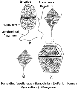
It include all unicellular (or acellular) eukaryotic animals. These are most primitive organisms considered as animals because of heterotrophic nutrition and motility. About 50,000 species (30,000 present and 20,000 extinct) are so far known. Brief history : Protozoans were first studied by Leeuwenhoek (1677). The name “Protozoa” was coined by Goldfuss (1817). The branch of their study is called Protozoology. General characters (1) Protozoans are the simple and primitive organisms. (2) They are free living or parasitic. (3) All the free living forms are aquatic. (4) They are asymmetrical or radially symmetrical or bilaterally symmetrical. (5) They are unicellular (acellular). (6) They have protoplasmic grade of organization. (7) Locomotion is effected by flagella, cilia or pseudopodia. (8) Nutrition is holophytic, holozoic, saprozoic or parasitic. (9) Digestion is intracellular. (10) Excretion and respiration occurs by diffusion. (11) In fresh water protozoans osmoregulation is carried out by the contractile vacuoles. (12) Encystment is a common phenomenon. (13) Reproduction occurs by asexual and sexual methods. Classification of Protozoans Protozoans are classified on the basis of locomotory organelles into following classes. Class 1. Rhizopoda or Sarcodina (1) There is no definite cell wall or pellicle. (2) There is no definite shape. (3) The locomotory organs are pseudopodia. (4) There is no permanent mouth or anus. (5) The contractile vacuoles are present in the fresh water forms. The rhizopoda has been divided into five orders. They are as Lobosa, Filosa, Foraminifera, Heliozoa and Radiolaria. Examples : Amoeba, Entamoeba histolytica, Entamoeba coli, Pelomyxa, Globigerina, Actinophryx.
  • In Arcella has an exoskeleton of tactin.
  • Giardia is called ‘Grand old man of intestine’.
  • Actinophryx is called ‘Sun animalcule’ as it resemble the sun.
Class 2. Flagellata or Mastigophora (1) The body is covered by a thin pellicle or cuticle. (2) The locomotory organs are flagella. (3) The contractile vacuoles are present in fresh water forms with accessory vacuoles. (4) Chloroplast are found in some forms. (5) They may be free living or parasitic. The class flagellata has been divided into eight orders. They are as Chrysomonadina, Cryptomonadina, Euglenoidea,  Phytomonadina, Dinoflagellata, Cystoflagellata, Protomonadina and Polymastigina. Examples : Chrysamoeba, Cryptomonas, Volvox, Chlamydomonas, Noctiluca, Mastigamoeba, Monal, Bado, Trypanosoma, Leishmania, Proterospongia etc.
  • Trychonymph (symbiotic) live in alimentary canal of termite that is digest to cellulose.
  • Noctiluca shows bioluminiscense due to luciferin protein. It is also called the ‘Fire of sea’.
Class 3. Sporozoa (1) They are exclusively endoparasitic. (2) The body is covered by pellicle. (3) Reproduction takes place by spore formation. The class is divided into two sub-classes, namely, Telosporidia and Neosporidia. Sub-class (i) Telosporidia (1) The spores do not contain polar capsules or filaments. (2) The life history ends with the formation of spores. (3) The spore cases are simple and contain many spores. Examples : Monocystis, Gregarina, Isopora, Eimeria, Plasmodium, Babesia etc.
  • Babesia causes the ‘Taxas cattle fever’ in animals. This disease also called ‘Red water fever’ or Haemoglobin uric fever.
  • Monocystis is more...


You need to login to perform this action.
You will be redirected in 3 sec spinner