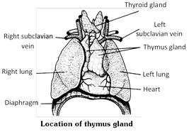
 Function of thymus glands
(1) Thymus is haemopoietic, as well as, an endocrine gland. Thymus is the "seedbed" of "thymic lymphocytes (T-lymphocytes). Certain "stem cells", originating in yolk sac and liver in early embryo, but only in bone marrow in late embryo, migrate into the thymus and proliferate to form a large number of lymphocytes.
(2) The major function of thymus is to secrete thymosin hormone, thymic humoral factor (THF), thymic factor (TF), thymopoietin. These compounds induce, not only the proliferation of lymphocytes, but also their differentiation into a variety of clones differently specialized to destroy different specific categories of antigens and pathogens likely to get into the body. This is called maturation of lymphocytes.
(3) As is clear from above account, thymus is essential in neonatal (newly born) infant and postnatal child for normal development of lymphoid organs and cellular immunity. That is why, the thymus, small at birth, progressively grows in size about three or four-folds upto about the age of puberty. By this time lymphoid organs and tissues are well-developed. The thymus, therefore, starts gradually diminishing in size and its tissue is progressively infiltrated by yellowish adipose tissue. This is known as the "immunity theory of ageing". By the old age, the thymus is reduced to quite a thin, yet functional chord of tissue.
Function of thymus glands
(1) Thymus is haemopoietic, as well as, an endocrine gland. Thymus is the "seedbed" of "thymic lymphocytes (T-lymphocytes). Certain "stem cells", originating in yolk sac and liver in early embryo, but only in bone marrow in late embryo, migrate into the thymus and proliferate to form a large number of lymphocytes.
(2) The major function of thymus is to secrete thymosin hormone, thymic humoral factor (THF), thymic factor (TF), thymopoietin. These compounds induce, not only the proliferation of lymphocytes, but also their differentiation into a variety of clones differently specialized to destroy different specific categories of antigens and pathogens likely to get into the body. This is called maturation of lymphocytes.
(3) As is clear from above account, thymus is essential in neonatal (newly born) infant and postnatal child for normal development of lymphoid organs and cellular immunity. That is why, the thymus, small at birth, progressively grows in size about three or four-folds upto about the age of puberty. By this time lymphoid organs and tissues are well-developed. The thymus, therefore, starts gradually diminishing in size and its tissue is progressively infiltrated by yellowish adipose tissue. This is known as the "immunity theory of ageing". By the old age, the thymus is reduced to quite a thin, yet functional chord of tissue. 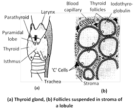 Synthesis and storage of iodothyroglobulin : Synthesis of a glycoprotein thyroglobulin (TGB) - occurs continuosly in the follicular cells under genic control. The cells keep extruding thyroglobulin in follicular cavity by exocytosis. Each molecule of thyroglobulin contains about 500 amino acid momoners of which 123 monomers are of tyrosine at fixed places. Soon as the molecules of iodine and thyroglobulin come out of follicular cells, these interact in such a way that 15 tyrosine monomers of each thyroglubulin molecule at fixed places become iodinated. Certain tyrosine monomers bind with single atoms of iodine, forming monoiodotyrosine (MIT or \[{{T}_{1}}\]). Other tyrosine monomers bind with two atoms of iodine, forming diiodotyrosine (DIT or \[{{T}_{2}}\]). This is called organification of thyroglobulin. Molecules of iodothyroglobulin keep accumulating in follicular cavity, forming the jelly-like colloid. Within the colloid, molecules of iodothyroglobulin undergo conformational changes and may even interact with each other. This results in a coupling of most of the iodinated tyrosine monomers in pairs. This more...
 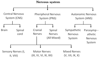 Central nervous system (CNS)
In all the vertebrates including man, CNS is dorsal, hollow and non-ganglionated while in invertebrates when present, it is ventral, solid, double and ganglionated. CNS is formed of two parts :
(1) Brain - Upper and broader part lying in the head.
(2) Spinal cord - Lower, long and narrow part running from beginning of neck to trunk. CNS is covered by 3 meninges and its wall has two type of matter.
Types of matter : CNS of vertebrates is formed of two types of matter –
(i) Grey matter : It is formed of cell-bodies, non-medullated nerve fibres, neuroglea, dendrites of association neurons and motor neurons.
(ii) White matter : It is formed of medullated nerve fibres or myelinated axon of motor and sensory neurons, which appear white due to presence of medullary sheath.
Meninges : The meninges are connective tissue membranes which surround the brain and spinal cord of CNS. In the fishes, there is only one meninx called meninx primitiva (piamater). In amphibians, reptiles and birds, the brain is covered by two meninges or membranes : inner pia-arachnoid and outer duramater. In mammals, CNS is covered by three meninges or membranes or cranial meninges. Brain meninges are continuous with spinal meninges
Central nervous system (CNS)
In all the vertebrates including man, CNS is dorsal, hollow and non-ganglionated while in invertebrates when present, it is ventral, solid, double and ganglionated. CNS is formed of two parts :
(1) Brain - Upper and broader part lying in the head.
(2) Spinal cord - Lower, long and narrow part running from beginning of neck to trunk. CNS is covered by 3 meninges and its wall has two type of matter.
Types of matter : CNS of vertebrates is formed of two types of matter –
(i) Grey matter : It is formed of cell-bodies, non-medullated nerve fibres, neuroglea, dendrites of association neurons and motor neurons.
(ii) White matter : It is formed of medullated nerve fibres or myelinated axon of motor and sensory neurons, which appear white due to presence of medullary sheath.
Meninges : The meninges are connective tissue membranes which surround the brain and spinal cord of CNS. In the fishes, there is only one meninx called meninx primitiva (piamater). In amphibians, reptiles and birds, the brain is covered by two meninges or membranes : inner pia-arachnoid and outer duramater. In mammals, CNS is covered by three meninges or membranes or cranial meninges. Brain meninges are continuous with spinal meninges
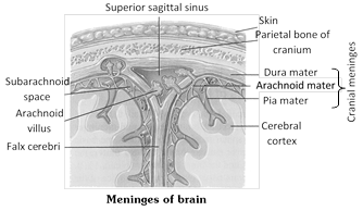 The three layers of cranial meninges in order from superficial to deeper duramater, arachnoid and piamater. Duramater is nonvascular, tough made up of fibrous connective tissue. Arachnoid mater made up of reticular connective tissue with collagen and elastin fiber, while innermost vascular piamater (nutritive) made up of loose aerolar connective tissue. Between dura and arachnoid mater presence of sub dural space (no CSF in mammals here), between Arachnoid and piamater presence of sub-arachnoid space (with CSF in mammals, CSF also found in ventricles and central canal). Between duramater and periosteum presence of epidural space. An extension of duramater between two cerebral hemispheres called falx cerebri. Tentorium, an extension of duramater between cerebrum and cerebellum.
Cerebrospinal fluid : All the ventricles of the brain, central canal of spinal cord are continuous and lined by a columnar, ciliated epithelium, the ependyma. They contain lymph-like extracellular fluid called the cerebrospinal fluid (C.S.F.). This fluid is secreted by the choroid plexuses by filtration of blood. The choroid plexuses consist of loose connective tissue of pia mater covered internally by a simple cuboidal epithelium of secretory (glandular) nature. The cerebrospinal fluid slowly flows toward the fourth ventricle by secretion pressure and passes into the spinal cord. Some fluid escapes into the subarachnoid spaces through three pores a median aperture (of magendie) and a paired lateral aperture (of Luschka) in the roof of the fourth ventricle in the medulla. From the subarachnoid spaces, the cerebrospinal fluid is transferred to the blood of the venous sinuses. Nervous tissue is more...
The three layers of cranial meninges in order from superficial to deeper duramater, arachnoid and piamater. Duramater is nonvascular, tough made up of fibrous connective tissue. Arachnoid mater made up of reticular connective tissue with collagen and elastin fiber, while innermost vascular piamater (nutritive) made up of loose aerolar connective tissue. Between dura and arachnoid mater presence of sub dural space (no CSF in mammals here), between Arachnoid and piamater presence of sub-arachnoid space (with CSF in mammals, CSF also found in ventricles and central canal). Between duramater and periosteum presence of epidural space. An extension of duramater between two cerebral hemispheres called falx cerebri. Tentorium, an extension of duramater between cerebrum and cerebellum.
Cerebrospinal fluid : All the ventricles of the brain, central canal of spinal cord are continuous and lined by a columnar, ciliated epithelium, the ependyma. They contain lymph-like extracellular fluid called the cerebrospinal fluid (C.S.F.). This fluid is secreted by the choroid plexuses by filtration of blood. The choroid plexuses consist of loose connective tissue of pia mater covered internally by a simple cuboidal epithelium of secretory (glandular) nature. The cerebrospinal fluid slowly flows toward the fourth ventricle by secretion pressure and passes into the spinal cord. Some fluid escapes into the subarachnoid spaces through three pores a median aperture (of magendie) and a paired lateral aperture (of Luschka) in the roof of the fourth ventricle in the medulla. From the subarachnoid spaces, the cerebrospinal fluid is transferred to the blood of the venous sinuses. Nervous tissue is more... 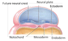
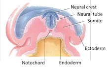
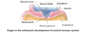 (1) Germinal layer : These are actively dividing cells lining the neural canal. They form the connective tissue lining of neural canal, called ependyma, and ventricles of brain.
(2) Mantle layer : It consists of embryonic neurons or nematoblasts, forming the gray matter.
(3) Marginal layer : It consists of nerve fibres, mostly surrounded by fatty myelin sheaths, and forms the white matter. Neurons and fibres are supported by a special connective tissue of ectodermal origin, the neuroglea, cells of which become increasingly abundant and diversified in higher vertebrates.
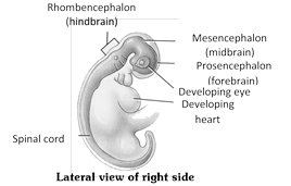
Development of brain
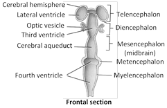
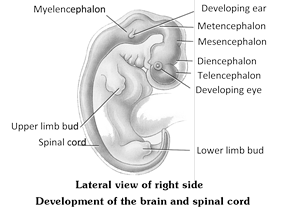
The anterior end of embryonic neural tube is already enlarged forming the embryonic brain, called encephalon. By differential growth and two constrictions, it is divided into a linear series of three primary cerebral vesicles, termed the forebrain, midbrain and hindbrain. These give rise to the three major divisions of the adult brain - (1) prosencephalon (forebrain), (2) mesencephalon (midbrain), and (3) rhombencephalon (hindbrain). These further become subdivided into 5 subdivisions. Prosencephalon divides into an anterior telencephalon and posterior diencephalon; the mesencephalon remain unchanged. The rhombencephalon divides into an anterior metencephalon and a posterior myelencephalon. Ultimately, telencephalon develops into cerebral hemisphere and basal ganglia and houses lateral ventricle. Diencephalon develops into thalamus, hypothalamus, and pineal gland and houses the third ventricle. Mesencephalon develops into mid brain and houses cerebral aqueduct. Metencephalon develops into pons and cerebellum; and myelencephalon develops into medulla oblongata, houses 4th ventricle. The area of neural tube inferior to myelencephalon gives rise to spinal cord.
(1) Germinal layer : These are actively dividing cells lining the neural canal. They form the connective tissue lining of neural canal, called ependyma, and ventricles of brain.
(2) Mantle layer : It consists of embryonic neurons or nematoblasts, forming the gray matter.
(3) Marginal layer : It consists of nerve fibres, mostly surrounded by fatty myelin sheaths, and forms the white matter. Neurons and fibres are supported by a special connective tissue of ectodermal origin, the neuroglea, cells of which become increasingly abundant and diversified in higher vertebrates.
Development of brain
The anterior end of embryonic neural tube is already enlarged forming the embryonic brain, called encephalon. By differential growth and two constrictions, it is divided into a linear series of three primary cerebral vesicles, termed the forebrain, midbrain and hindbrain. These give rise to the three major divisions of the adult brain - (1) prosencephalon (forebrain), (2) mesencephalon (midbrain), and (3) rhombencephalon (hindbrain). These further become subdivided into 5 subdivisions. Prosencephalon divides into an anterior telencephalon and posterior diencephalon; the mesencephalon remain unchanged. The rhombencephalon divides into an anterior metencephalon and a posterior myelencephalon. Ultimately, telencephalon develops into cerebral hemisphere and basal ganglia and houses lateral ventricle. Diencephalon develops into thalamus, hypothalamus, and pineal gland and houses the third ventricle. Mesencephalon develops into mid brain and houses cerebral aqueduct. Metencephalon develops into pons and cerebellum; and myelencephalon develops into medulla oblongata, houses 4th ventricle. The area of neural tube inferior to myelencephalon gives rise to spinal cord.

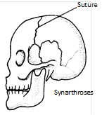 (iii) Syndesmosis : It is type of fibrous joint with more fibrous tissue than sutures. e.g., distal articulation between Tibia and fibula.
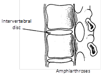
(2) Imperfect joints (Amphiarthroses) slightly movable : Joints in which syanovial cavity is absent. Permit a small amount of movement. Fibrocartilage is placed between the bones. These are cartilaginous joints e.g., Pubic symphysis, between bodies of the vertebrae, between the manubrium and the body of sternum, sacroilliac joint in frog.
(iii) Syndesmosis : It is type of fibrous joint with more fibrous tissue than sutures. e.g., distal articulation between Tibia and fibula.
(2) Imperfect joints (Amphiarthroses) slightly movable : Joints in which syanovial cavity is absent. Permit a small amount of movement. Fibrocartilage is placed between the bones. These are cartilaginous joints e.g., Pubic symphysis, between bodies of the vertebrae, between the manubrium and the body of sternum, sacroilliac joint in frog.
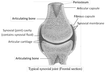
 (3) Perfect joints (Diarthroses) freely movable : Syanovial cavity and ligaments are present. These are typical joints having articulate surface and syanovial capsule. Syanovial fluid act as a grease in the joint e.g., Joints of elbow, ankle, wrist, hip, knee. Articular cartilage covers the surface of articular bones. Articular cartilage of synovial joint is hyaline cartilage. Synovial joints are surrounded by tubular articular capsule. The articular capsule consists of two layers, outer fibrous capsule and inner synovial membrane. The synovial membrane secretes synovial fluid which lubricates and provides nourishment to articular cartilage. In old age stiffness of joints is due to the decrease in synovial fluid.
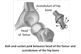
 (i) Ball and socket joint : Also known as enarthroses. Ball of one bone articulate in socket of another bone. e.g., head of humerus and glenoid cavity of pectoral girdle, femur and acetabulum of pelvic girdle, joint between incus and stapes.
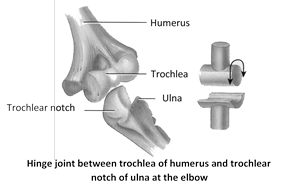
 (ii) Hinge joint : Also known as gingulum. Movement is possible in one direction only. e.g., Joint of malleus and incus, knee joint, elbow joint, articulation joint of lower jaw, joint of phalanges of digits.
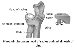
 (iii) Pivot joint : Also known as rotatoria and helps in turning movement. One bone is fixed and second articulate. e.g., Atlas and axial of skull rotate with axis vertebra also known as atlanto axial joint.
 (iv) Gliding joint : Also known as arthrodial, limited movement in all more...
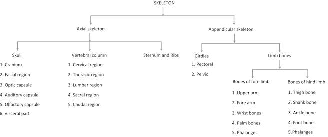
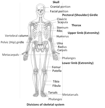 Axial skeleton (Human)
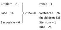
It occupies the body's main longitudinal axis. It includes four structure : skull in the head, vertebral column in the neck, trunk and tail if present, sternum and ribs in the thorax. It form the upright axis of body and includes 80 (87 in children) bones are as follows in man -
Axial skeleton (Human)
It occupies the body's main longitudinal axis. It includes four structure : skull in the head, vertebral column in the neck, trunk and tail if present, sternum and ribs in the thorax. It form the upright axis of body and includes 80 (87 in children) bones are as follows in man -
 (1) Skull (General structure) : It is anterior most axial skeleton. It is divisible into two main parts –
(i) Chondrocranium (ii) Splanchnocranium
(i) Chondrocranium : Chondrocranium is formed by (a) brain box or cranium proper and (b) two sense capsules - Orbit or optic capsule (eye) and auditory or otic capsule (ear).
(a) Cranium proper : It is a strong and firm bony box with a helmet-like covering over the brain, called vault of skull, and a relatively thicker and stronger floor of base upon which the brain rests. Its cavity is called cranial cavity. Size of cranial cavity averages 1475 cubic centimetres \[(c{{m}^{3}})\] in adult men. At about the middle of the floor of cranium, there is a large opening of cranial cavity called foramen magnum. The brain is connected to spinal cord at this foramen. Cranium proper of mammal has following distinct zone -
(1) Skull (General structure) : It is anterior most axial skeleton. It is divisible into two main parts –
(i) Chondrocranium (ii) Splanchnocranium
(i) Chondrocranium : Chondrocranium is formed by (a) brain box or cranium proper and (b) two sense capsules - Orbit or optic capsule (eye) and auditory or otic capsule (ear).
(a) Cranium proper : It is a strong and firm bony box with a helmet-like covering over the brain, called vault of skull, and a relatively thicker and stronger floor of base upon which the brain rests. Its cavity is called cranial cavity. Size of cranial cavity averages 1475 cubic centimetres \[(c{{m}^{3}})\] in adult men. At about the middle of the floor of cranium, there is a large opening of cranial cavity called foramen magnum. The brain is connected to spinal cord at this foramen. Cranium proper of mammal has following distinct zone -


You need to login to perform this action.
You will be redirected in
3 sec

