Current Affairs 11th Class

Dauclax, (1883) introduced the nomenclature of enzyme. Usually enzyme names end in suffix-ase to the name of substrate e.g., Lactase acts on lactose, maltase act on maltose, amylase on amylose, sucrase on sucrose, protease on proteins, lipase on lipids and cellulase on cellulose. Sometimes arbitrary names are also popular e.g., Pepsin, Trypsin and Ptylin etc. Few names have been assigned as the basis of the source from which they are extracted e.g., Papain from papaya, bromelain from pineapple (family Bromeliaceae). Enzymes can also be named by adding suffix-ase to the nature of chemical reaction also e.g., Oxidase, dehydrogenase, catalase, DNA polymerase. Modern names are given after chemical action. They are more systamatic, informative but slightly longer. e.g., ATP : D-glucose phosphotransferase. Common simpler names used at the place of systematic names called trivial names. According to older classification : The older classification of enzymes is based on the basis of reactions which they catalyse. Many earlier authors have classified enzymes into two groups : (1) Hydrolysing enzyme : The hydrolysing enzymes of hydrolases catalyse reactions in which complex organic compounds are broken into simpler compounds with the addition of water. Depending upon the substrate hydrolysing enzymes are : Carbohydrases : Most of the polysaccharides, disaccharides or small oligosaccharides are hydrolysed to simpler compounds, e.g., hexoses or pentoses under the influence of these enzymes. Lactase on lactose to form glucose to galactose, sucrase/invertase on sucrose to form glucose and fructose, amylase or diastase on starch to form maltose, maltase on maltose to form glucose, cellulase on cellulose to produce glucose. Easterases : These enzymes catalyse the hydrolysis of substances containing easter linkage, e.g., fat, pectin, etc. into an alcoholic and an acidic compound. \[Fat\xrightarrow{Lipase}Glycerol+Fatty\text{ }acid\] \[Phosphoric\,acid\,easters\xrightarrow{Phosphatase}\] \[Phosphoric\text{ }acid+Other\text{ }compounds\] Proteolytic enzymes : The hydrolysis of proteins into peptones, polypeptides and amino acids is catalysed by these enzymes \[\Pr otein\xrightarrow{Pep\sin }Peptones\] \[Polypeptides\xrightarrow{Peptidases}A\min o\text{ }acids\] Amidases : They hydrolyse amides into ammonia and acids. \[Urea\xrightarrow{Urease}Ammonia+Carbon\text{ }dioxide\] \[Asparagine\xrightarrow{Asparaginase}Ammonia+Aspartic\text{ }acid\] (2) Desmolysing enzymes :  Most of the desmolysing enzymes are the enzymes of respiration e.g., oxidases, dehydrogenases, (concerned with transfer of electrons), transaminases carboxylases etc. According to IUB system to classification : In 1961 the Commission on enzymes set up by the 'International Union of Biochemistry' (IUB) framed certain rules of their nomenclature and classification. According to IUB system of classification the major points are : • Reactions (and enzymes catalyzing them) are divided into 6 major classes each with 4-13 subclasses. • The enzyme name has two parts-first name is of substrate. The second ending in ase indicates type of reaction. • The enzyme has a systematic code No. (Enzyme code/Enzyme Commission). The first digit denotes the class, the second sub-class, the third sub-sub-class and the fourth one is for the particular enzyme name. Thus, E.C. 2.7.1.1 denotes class 2 (Transferases)-subclass 7 (transfer of phosphate) sub-sub-class 1 (an alcohol functions as phosphate acceptor). The 4th digit indicates hexokinase. Major classes of enzymes are as follows : (i) Oxidoreductases : These enzymes catalyse oxidation reduction more...

Molecular weight : Enzymatic proteins are substances of high molecular weight. Bacterial ferredoxin one of the smaller enzymes has molecular weight of 6,000, where as pyruvic dehydrogenase one of the largest-has a molecular weight of 4600000. Amphoteric nature : Each molecule of enzyme possess numerous groups which yield H+ in slightly alkaline solutions and groups which yield OH- ions in slightly acidic solutions. Unlike many other substances, therefore, the enzymatic protein is amphoteric, i.e., capable of ionizing either as an acid or as a base depending upon the acidity of the external solution. Colloidal nature : All enzymes are colloidal in nature  and thus provide large surface area for reaction to take place. They posses extremely low rates of diffusion and form colloidal system in water. Specificity of enzyme : Most of the enzymes are highly specific in their action. A single enzyme will generally catalyze only a single substrate or a group of closely related substrates. The active site possess a particular binding site which complexes only with specific substrate. Thus, only a suitable substrate fulfils the requirements of active site and closely fixes with it. Heat specificity : The enzymes are thermolabile i.e., heat sensitive. They function best at an optimum temperature \[(20{}^\circ C-40{}^\circ C).\] Their activity decrease with decrease as well as increase in temperature and stops at \[0{}^\circ C\] and above \[80{}^\circ C.\] Catalytic properties : Enzymes are active in extremely small amounts, e.g., one molecule of invertase can effectively hydrolyze 1,000,000 times its own weight of sucrose. One molecule of catalase is able to catalyze conversion of 5,000,000 molecules of hydrogen peroxide. Reversibility of reaction : The enzyme-controlled reactions are reversible. The enzymes affect only the rate of biochemical reactions, not the direction. e.g., Lipase can catalyse splitting of fat into fatty acids and glycerol as well as synthesis of fatty acids and glycerol into fats. \[FatGlycerol+Fatty\text{ }acid\] pH sensitivity : The enzymes show maximum activity at an optimum pH is \[6-7.05\,\,(7\pm 1.05).\] Their activity slows with decrease and increase in pH till it stops. Each enzyme has its own different favourable pH value. High efficiency : The effectiveness of an enzymatic reaction is expressed in terms of its turn over number or catalytic centre activity means number of substrate molecules on which one enzymes molecules acts in one minute. Turn over number depends on the number of active sites of an enzyme. An active site is an area of the enzyme which is capable of attracting and holding particular substrate molecules by its specific charge, size and shape so as to allow the chemical change, Enzymes show 3-D structure. R (alkyl) groups of amino acids from active sites during folding polypeptide chains. Usually 3-12 amino acids form an active site. Highest turn over number is of carbonic anhydrase (36 million/min or 600000 per second) and lowest is of lysozymes (30/min or 0.5 per second). So carbonic anhydrase is fastest enzyme. The lowest turn over number is of lysozymes.

All enzymes are produced in the living cells. About 3,000 enzymes have recorded. These are of two types with regard to the site where they act as : Intracellular enzymes :  Most of the enzymes remain and function inside the cells, They are called the intracellular enzymes or endoenzymes. Some of these enzymes are found in cytoplasmic matrix. Certain enzymes are bound to ribosomes, mitochondria and chloroplast etc. Extracellular enzymes : Certain enzymes leave the cells and function outside them. They are called the extracellular enzymes or exoenzymes. They mainly include the digestive enzymes. e.g., salivary amylase, gastric pepsin, lysozyme present in tears and nasal secretion. Rennet tablets with enzyme renin from calf's stomach are widely used to coagulate protein caseinogen for cheese (casein) formation.

Zymogens or (Enzyme Precursors) : Certain enzymes are produced by the living cells in an inactive (non-functional) form. They are called the zymogens or proenzymes. It is then converted, usually by proteolysis (hydrolysis of the protein), to the active form when it has reached the site of its activity. Pepsinogen and trypsinogen are zymogens produced by gastric glands and pancreas respectively. They are necessary to life because they degrade dietary proteins into amino acids that are used by the cell. Pepsinogen is changed to active pepsin by hydrogen ions in the stomach. Trypsinogen is activated to trypsin by an enzyme enterokinase in the small intestine. Once small amount of pepsin or trypsin is formed, it itself catalyzes the activation of remaining proenzyme. This process is called autocatalytic reaction, or autocatalysis. Isoenzymes (Isozymes) : There are certain enzymes which have slightly different molecular structure but performing the same catalytic function. Such enzymes are called isoenzymes or simply isozymes. Isoenzyme of an enzyme differ from each another in their amino acid sequence, molecular weight, immunological and electrophoretic behaviours. Hence, they can be separated by electrophoresis. More than 100 enzymes are known to have isoenzyme. A good example of isoenzyme is lactic dehydrogenase (LDH). It catalyzes change of pyruvate to lactate. There are five LDH isoenzymes in muscles of heart. Alcohol dehydrogenase has four isoenzyme in maize. a-amylase (wheat endosperm) has 16 isoenzymes. Inducible enzymes : An enzyme which is synthesized only in the presence of its substrate (inducer) is called inducible enzyme e.g., \[\beta -\]galactosidase. Constitutive enzymes (House keeping enzyme) : The enzyme which are found in constant amounts under different growth conditions (regardless of its metabolic states) are called constitutive enzyme e.g., enzymes of sugar breakdown i.e., glycolysis. Repressible enzymes : The presence of a specific substance may inhibit continued production of specific enzyme (enzyme repressor) e.g., glucokinase. Ribozymes : Study of post transcriptional processing of RNA molecules has led to the most exciting discovery of the existence of some catalytic RNA molecules which have been called as RNA enzymes or ribozymes. All enzymes are not proteins as confirmed by Cech (1981) and Altman (1983). Ribozyme and RNAase-P are two non protein enzyme where RNA acts as catalyst. Ribozyme was reported from Tetrahymens (a protozoans) by Cech. The substrate for ribozyme is usually an RNA molecule. RNAase-P (Ribonuclease) was discovered by Altman. Peptidyl transferase is also a non-proteinaceous enzyme, discovered by Noller. Michaelis constant : Michaelis and Menten (1913) introduced a constant \[{{K}_{m}}\] (Michaelis constant). It is a mathematical derivative or constant which indicates the substrate concentration at which the chemical reaction catalysed by an enzyme attains half its maximum velocity \[({{V}_{\max }}).\] \[{{K}_{m}}\]indicates affinity of the enzyme for its substrate.                 \[{{K}_{m}}=\frac{1}{2}{{V}_{\max }}\] \[{{K}_{m}}\]value differs from substrate to substrate because different enzymes differ in their affinity towards different substrates. A high \[{{K}_{m}}\] indicates low affinity while a low \[{{K}_{m}}\] shows strong affinity. Protease acts on different proteins. So it's \[{{K}_{m}}\] value will differ from protein to protein.   more...

Systematic position                 Phylum          -    Chordata                 Sub phylum -    Vertebrata or Craniata                 Class               -    Amphibia                 Order             -    Salientia or Anura                 Genus            -    Rana                 Species          -    tigrina   Habitat Rana tigrina is the most widely distributed species in Northern India. Generally frogs are found in ponds, tank, pools, ditches, etc. However, they may leave their aquatic habitat to come on land to hunt for their prey, which are mostly insects. Habits (1) Locomotion : (a) Jumping and leaping, (b) Swimming.  Absence of neck is helpful in swimming in water and jumping on land. (2) Feeding : The adult frog is carnivorous. Tadpole (larva of frog) is herbivorous. (3) Croaking : The male frog croaks louder than the females because of the presence of two vocal sacs in male frog. The vocal sacs act as resonators. The croaking is mating call to attract the female frog. (4) Hibernation (Winter sleep) : During hibernation frog respires through skin (cutaneous respiration) only. (5) Aestivation (Summer sleep) : During this period frog takes rest and recuperates its energy. (6) Protective Colouration : The frog is capable of changing its body colour with the change in its surroundings. It can not only avoid its enemies but can catch its prey unnoticed. (7) Breeding : The male frog jumps on the female frog and holds her tightly with the help of his fore-limbs. Gripping of the female by the male is also very much aided by the presence of nuptial pads. This sexual embrace is called the amplexus. Fertilization is external. During development, a fish like tailed tadpole is produced, which respires with the help of gills and feeds upon vegetable matter. (8) Moulting : The frog sheds off almost once a month its skin during its active life in the form of small casting. This phenomenon is known as moulting. External morphology The dorsal surface of frog is spotted olive green and ventral pale yellow; this protective colouration help to camoflage, i.e. escape the detection by enemies. Body division : The body of frog has two main divisions head and trunk; absence of neck and tail helps both in jumping on land and swimming in water. (1) Head : Head is anterior flattened triangular part with, a wide transverse terminal mouth, a pair of small dorsal external nares, two dorso-lateral eyes, a mid-dorsal light coloured brow spot or third eye or pineal body and obliquely placed pigmented circular tympanum or ear drum. Eyes are provided with nictitating membrane for protection. (2) Trunk : Trunk is the large, oval, flattened main part of the body. It is differentiated into hard anterior thorax and soft posterior abdomen. The trunk is provided with a pair of fore and hind limbs. The fore limbs are shorter and stouter, end in four digits. The more...

Systematic position                 Phylum          -  Chordata                 Subphylum   -  Vertebrata or Craniata                 Class                -  Mammalia                 Order              -  Rodentia                 Family             -  Muridal                 Genus            -  Rattus                 Species          -  rattus (Black Rat) Habits and Habitat They are the common house rats which are cosmopolitan in distribution and found all over the world. They are herbivorous, fossorial and nocturnal animals and undergo hibernation. They show sexual dimorphism. They are prolific breeders. Fertilization is internal. The time interval between fertilization and birth (gestation) is about 22 to 23 days. They are completely grown at six to eight months of age. The rat breeds more than four times in a year producing 6-8 young ones in each litter. Newly born young ones are blind, deaf and without hairs. The mother feeds the young ones on milk. Average age of a rat is 3 years. External morphology Their body is covered with hairs. The body is divisible into head, neck, trunk and tail. Head : Head is broader posteriorly and tapers anteriorly as a naked terminal muzzle or snout. A pair of nostrils, shaped like inverted commas, is present above the mouth opening, which leads into nasal passages. Below the nostrils is the cleft upper lip, which exposes the two upper incisors. On the lateral sides of the head are large, paired bulging eyes. Eyelids have very fine and short eyelashes; the nictitating membrane is reduced. The head bears a pair of external ear or pinna at its posteriolateral position. The mouth is sub-terminal and located beneath the nostrils and remains guarded by upper and lower lips. Long, stiff, bristle-like hair known as pili lactiles or vibrissae are present on both sides of nostrils. They help the animal in measuring width of area through which the animal is to pass even in perfect darkness. Neck : It is a short connective between head and trunk. With the help of neck the animal can bend its head in different directions. Trunk : It is depressed fusiform major part of the body which has two parts-anterior narrow but stouter thorax and posterior wider softer abdomen. The ventral surface of female bears 6 pairs of teats or nipples, three pectoral (thoracic) and three inguinal (abdominal). The trunk bears two pairs of limbs, two forelimbs and two hindlimbs. Forelimbs are smaller than the hindlimbs. Each limb is made up of proximal segment (stylopodium), middle segment (Zeugopodium) and distal segment (autopodium). Five digits are present in autopodium of each limb. The first digit is thumb or pollex, which is much reduced with a peculiarly flattened nail and two phalanges. Nail is keratinized structure occupying position above the distal phalanx of each digit. Typical walking pads, the tori are present on the tips of digits, palm and at the base of palm. These are also more...

It connects and supports all the other tissues, the intercellular element predominating. The cellular element is usually scanty. In function this tissue may be mechanical, nutritive and defensive. It is a tissue made up of matrix (abundant intercellular substance or ground substance) and living cells that connects and support different tissues. All connective tissues in the body are formed by mesoderm. Structure There are large intercellular spaces between the cells. Intercellular spaces are filled with large amount of extracellular materials formed of insoluble protein fibres lying in an amorphous, transparent ground substance called matrix. Ageing of an animal body is associated with deterioration in its connective tissues. Functions (1) Their chief function is to bind other tissues together in the organs. (2) Certain connective tissues such as adipose tissues store fat. (3) Skeletal connective tissues like bones and cartilages provide the body with a supporting skeletal frame work. (4) Fluid connective tissues such as blood and lymph transport various materials in the body. (5) Plasma cells synthesize antibodies, viz., macrophages. Lymphocytes ingest cell debris, harmful bacteria and foreign matter. Thus these cells of connective tissues are protective in function. (6) The jelly-like ground substance of connective tissues acts as shock absorber around some organs such as eye balls and kidneys. (7) The bone marrow produces blood cells. (8) Areolar tissue acts as packing material in various organs. (9) Collagen fibres of connective tissue help in repair of injured tissues. Types of connective tissues Connective tissue proper possess soft viscous semisolid or semi-fluid matrix. It is divided into following types : (1) Areolar Tissue : Areolar tissue is loose connective tissue, possess transparent gelatinous, highly vascular and sticky matrix which have variety of cells and fibres. It allows movement of part connected by it (Muscle and their compound). Areolar tissue mainly consist of different types of cells and fibres.      
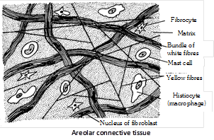
(i) Cells of areolar tissue : It has following types – Fibroblast : It is most abundant cells, produces fibres, called as fibroblasts in their young active phase and fibrocytes when old and inactive. It synthesize proteins (Collagen, elastin and reticulin). These are undifferentiated mesenchyme stem cells, capable to give rise other cells of connective tissue. Collagen and elastin are formed by fibroblasts. 
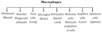
Histiocytes or Macrophages or Clasmatocytes  : These are polymorphic cells. These are amoeboid cells and these are main phagocytes of connective tissue. They are having most active lysosomes and phagocytise dead cells and pathogens. Macrophages remove the dead cells and damaged cells and clean the body so called scavenger cell. All types of macrophages take part in phagocytosis.      Reticular cells : Present only in the reticular tissue and stellate in appearance. Infact they are modified fibroblast producing reticular fibres. 
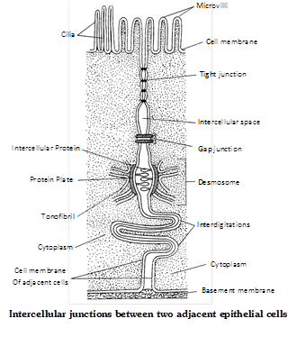
Mast cells : Mast cells were discovered by Paul Echrlich. It is large, irregular ovoid cells found in more...

An epithelium is a tissue composed of one or more layers of cells that cover the body surface and lines its various cavities. It serves for protection, secretion and excretion. The word ‘epithelium’ (G. epi = upon, thele = nipple) was introduced by Ruysch. They are located on the outer surfaces of organs, including the skin. They form the linings of tracts, cavities and vessels. Epithelial tissue evolved first in animal kingdom. It originate from all the three primary germ layers. Structure Cells are arranged in one or more layers, cells are compactly arranged and there is no inter cellular matrix between them. Neighbouring cells are held together by intercellular junctional complexes like desmosomes, tight junctions, interdigitations etc. The cells of lowermost layers always rest on a non living basement membrane or basal lamina. Basement membrane is made up of no cell product of epithelial tissue. It is formed of mucopolysaccharides, glycoprotein and collagen or reticular fibres. Blood vessels are absent in the epithelial tissues. However, nerve endings may penetrate the epithelium. It posses very high capacity of renewal (mitotic cell division). The following types of modifications and junctions are found in the plasma membrane of adjacent epithelial cells to keep the cells together.       Microvilli : It is simple and minute cytoplasmic processes arising from free exposed surfaces of the cell. They absorb material. e.g. Intestine. Stereocilia : It is non-motile cytoplasmic processes. e.g. Epididymis, vas deference. Kinocilia : It is contractile motile fibrous processes arising from basal granules. e.g. Oviduct, Fallopian tube. Tight junctions  (Zona occludens) : At certain places the plasma membranes of adjacent cells are tightly packed or even fused together. e.g. Brain. Desmosomes : Desmosome is present in epithelial tissue. They consist of thickened area and several fine tonofibrils extending from each plasma membrane into cytoplasm of respective cells. Macula adherens is a kind of desmosome. e.g. Vagina, Urinary bladder. Gap junction : At place, the adjacent cells form ion-rich gap junctions for intercellular communication and chemical exchange. These junctions probably do not provide physical support. Interdigitations : These are interwoven finger-like processes of plasma membranes of adjacent cells. Intercellular bridges : These are minute projections that arise from adjacent cell membranes. The intercellular bridges make contact with one another.     Functions Epithelial tissues have a wide spread distribution throughout the body and serve several important functions – (1) Generalized protection is the most important function of membranous epithelium. It is the relatively tough and impermeable epithelial covering of the skin that protects the body from mechanical and chemical injury and also from invading bacteria and other disease causing micro-organisms. (2) Epithelial structures specialized for sensory functions are found in the skin, nose, eye and ear. (3) Glandular epithelium is specialized for secretory activity, secretory products include hormones, mucous, digestive juices and sweat. (4) The more...

Glandular epithelium are specialized for secretory activity. A cell, tissue or organ which secretes a useful chemical material is known as gland. Glands are made up of cuboidal epithelial cells which are more secretory. All glands arise as folding of epithelia. The golgi body in gland cells are larger and more secretory. Most of the glands of body are merocrine types. It originate from all three germinal layers. (ecto, meso and endoderm). Liver is the largest gland of the body and lined by glandular epithelium. Types of glands (1) Unicellular gland : It  consist of unicellular gland cells which are called as goblet cells or chalice cells. They secrete mucous and found in mucosa of intestine and stomach. Mucous lubricates the food for easy peristalsis. Their life span is about \[2\,\,\,\,3\] days. (2) Multicellular gland : It consist of many cells and are generally located in underlying connective tissue e.g. gastric and intestinal glands. (3) Exocrine gland : These are those glands which discharge their secretory products into ducts. It is also called ducted glands or glands of external secretion.  e.g. Salivary glands, Mammary glands and Tear glands. (4) Endocrine gland : It is often called ductless gland, because they discharge their secretory products (hormones) directly into the blood. e.g. Pituitary gland, thyroid, parathyroid and adrenal glands. (5) Heterocrine gland : These are those glands which are partly endocrine and partly exocrine in function. e.g. Pancreas. Structural classification of exocrine glands Multicellular exocrine glands are classified by structure, using the shape of their ducts and the complexity (branching) of their ducts system as distinguishing characteristics. Shape include tubular and alveolar (Sac like). Simple exocrine glands e.g. intestinal glands, mammalian sweat glands, cutaneous glands of frog etc. have only one duct leading to surface. Compound exocrine glands have two or more ducts e.g. liver, salivary glands etc.   
Type Example
Simple tubular Intestinal glands, crypts of Lieberkuhn in ileum.
Simple coiled tubular Sweat glands in man
more...
Contractility and motility (movement) are fundamental properties of protoplasm. That is why, all cells possess potential motility. Contraction for motility in the cells results essentially from the interaction of two contractile proteins, actin and myosin. These tissues are obviously responsible for movements of organs and locomotion of the body in response to stimuli. These develop from embryonic mesoderm except for those of the iris and ciliary body of eyes, which are ectodermal in origin. About 40% to 50% of our body mass is of muscles. The muscle cells are always elongated, slender and spindle-shaped, fibre-like cells, These are, therefore called muscle fibres. These possess large numbers of myofibrils formed of actin and myosin. Muscle cells lose capacity to divide, multiply and regenerate to a great extent. Study of muscle is called myology. Types of muscle are following – Striated or striped muscles Most muscles of body are striated. These generally bring about voluntary movements under conscious control of brain and, hence, called voluntary muscles. Most of these are inserted at both ends upon bones in different parts of the body depend upon these muscles. Hence, these are also called skeletal muscles. Movements of limbs and the body solely depend upon these muscles. Hence these are also called somatic muscles. These are also called phasic type of muscles, because contraction in these is rapid, but brief and fatigue occurs quickly. Fine structure of striated muscle fibres : Striated muscle fibres shows transverse striation in the form of regular alternate dark A (anisotropic) and light I (isotropic) bands. The ‘A’ band contains about \[120{AA}\] thick and \[1.8\,\,\mu \] long “myosin filaments”. The I band contains about \[60{AA}\] thick and \[1.0\,\,\mu \] long “actin filament” which are twice as many as myosin filaments. Each I band is divided into two equal halves by a thin, fibrous and transverse zig-zag partition, called ‘Z’ band (‘ Z’ disc) or Krause’s membrane. Each segment of a fibril between two adjacent ‘Z’ bands is called a sarcomere. It is \[2.3\,\,\mu \] long in uncontracted mammalian striated fibres. A slender transverse line, the ‘M’ or Hansen’s line is visible in middle of each ‘A’ band. The major, middle region of ‘A’ band is comparatively lighter, but its terminal parts appear darker. The middle lighter region is called ‘H’ zone. Due to the geomatric bonding pattern, the end of each myosin filament is, thus, encircled by the ends of six actin filaments (hexagon), while the end of each actin filaments is encircled by the ends of three myosin filaments (trigon). Ultrastructure of myofilaments : At the molecular level, each myosin filament is composed of about 500 thread-like myosin molecules. Three different kinds of proteins participate in the composition of actin filaments. The major part of an actin filament is a coiled double helical strand whose each arm is a linear polymer of small and globular molecules (monomers) actin protein. Another coiled double helical, but thiner, strand runs along the whole length of actin strand. Each arm of this strand more...


You need to login to perform this action.
You will be redirected in 3 sec spinner