Implantation
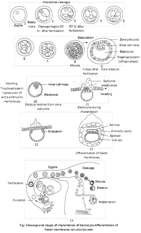
(i) Definition: The process of attachment of the blastocyst on the endometrium of the uterus is called implantation.
(ii) Period: Though the implantation may occur at any period between 6th and 10th day after the fertilization but generally it occurs on seventh day after fertilization.
(iii) Mechanism: First of all, the blastocyst is held closely against the uterine endometrial epithelium. The uterine capillaries and uterine wall in the immediate vicinity of the embryo become more permeable and a local stromal edema is developed. Soon the endometrium around the embryo shows the first sign of a decidual cell reaction (DCR) which involves:
(a) The epithelium becomes disrupted and the loosely packed fibroblast-like cells of the stoma are transformed into large rounded glycogen-filled cells.
(b) The area of contact becomes more vascular.
(c) The decidual cells form an “implantation chamber” around the embryo before the formation of a functional placenta.
(d) The tropho blast is developed from the superficial layer of the morula stage. Later, the trophoblast is lined by mesoderm to form the chorion which contributes to the placenta formation.
(e) Trophoblast of the chorion penetrates the uterine epithelium by both cytolytic and mechanical activity. The phagocytic activity of the trophoblastic cells through the decidual cells continues till it establishes intimate connection with the uterine blood vessels. The process of implantation is aided by proteolytic enzymes produced by the trophoblast. After implantation, endometrium undergoes many changes and forms decidua. It is differentiates into three parts such as : Decidua basalis present between the embryo and uterine myometrium, Decidua capsularis lies between the embryo and lumen of the uterus and Decidua parietalis is formed by the remaining part of decidua. The pattern of implantation of the blastocyst varies in different species, which are as follows
(1) Interstitial implantation: The blastocyst get burried into the endometrium e.g. human female, hedgehog, guinea pig, some bats and ape.
(2) Central implantation: The blastocyst remain the uterine cavity e.g. rabbit, cow, dog and monkey.
(3) Eccentric implantation: The blastocyst comes to lie in a uterine recess e.g. rats, mice.
(iv) Hormonal control of implantation
(a) Role of estrogens: These are a group of steroid hormones mainly secreted by follicular epithelial cells of Graafian follicle though these are also produced by adrenal cortex and placenta. These include b-estradiol, esterone, estriol etc. Out of which most important estrogen is b-estradiol. Secretion of estrogens is stimulated by FSH of anterior lobe of pituitary glands. These stimulate the uterine endometrial epithelium to enlarge, become more vascular and more glandular. The uterine glands become tortuous and cork-screw shaped. So the endometrium prepares itself for implantation. This stimulation by the estrogens on the uterus generally occurs on the 4th day of pregnancy.
(b) Progesterone: It is also a steriod hormone secreted by yellow-coloured endocrine gland, called corpus luteum, formed from empty Graafian folicle during more...
Neurulation and organogenesis
Post gastrulation involves two main process. Neurulation is process of laying the neural plate to form the nervous system. The establishment of the germ layers initiates the final phase of embryonic development, i.e., organogenesis. The latter involves differentiation and specialization of groups of cells in the individual germ layers. The cells of such groups change their form and give rise to morphologically recognizable tissues and organs of the new individual. The groups of differentiated cells separate from their germ layers in an orderly manner and with unique precision. Separation of the differentiated cell groups may occur by folding off from the germ layer or by migration of cells individually and aggregation at a new place. In this manner, the primordial cells of the germ layers gradually and accurately give rise to the tissues and organs of the offspring.
By four weeks after fertilization, the embryo has a simple heart, limb buds and eye rudiments. It also has a tail and pharyngeal pouches, the vestiges of its early vertebrate ancestors that disappear later in development. After the second month, the embryo is recognizable as a primate. From this stage on, the embryo is often called fetus.
Some important events in the human development
Time from fertilization
Stage/organs
Event
24 hours
Cleavage
Embryo is at two-cell stage
3 days
Modula
The module reaches uterus
7 days
Blastocyst
Implantation of blastocyst begins
2.5 weeks
Notochord
Notochord formed, differentiation of tissues that will give rise to heart, blood cells formed in yolk sac and chorionic.
3.5 weeks
Organ system
Neural tube formed, primordial eye and ear vesicle, pharyngeal pouches formed, liver bud differentiates, respiratory system and thyroid gland begin to develop, heart tube bends and begins to beat, blood vessels are formed.
4 weeks
Limb buds
Development and appearance of limb buds, brain forms three primary vesicles.
2 months
Muscles and gonads
Muscles differentiate, embryo capable of movement, gonads distinguishable as testes or ovaries, ossification of bones begins, cerebral cortex is differentiated, blood vessels assume final position.
3 months
Sex differentiation
By external examination sex can be determined, notochord degenerates, lymph glands develop.
Extra embryonic membrane
An aquatic embryo is surrounded by water, which protects the embryo, keep it moist, removes wastes and permits gas exchange. In land vertebrate (reptiles, birds and mammals), these functions are taken over by the extra embryonic membranes. These membranes are formed outside the embryo from the trophoblastic only in amniotes and perform specific function. Some of these membranes take part in the formation of placenta in mammals.
(i) Yolk sac: It is formed below the embryo. It contains fluid, not yolk. The yolk sac is a vestigial organ inherited from the oviparous reptilian ancestors. Yolk sac encloses by outer mesoderm and inner endodermal layer.
Function: It is mainly digestive in function. It also absorbs the dissolved yolk and passes it to developing embryo in reptiles, birds and prototherian. In human beings, it is vestigial. In human embryo it act as the site of blood cell formation until about the 6th week, when the liver takes over this role.
(ii) Amnion: It is formed above the embryo. It consist of outer mesoderm and inner ectoderm. The amnion and the fluid filled amniotic cavity it encloses, enlarge and nearly surround the embryo. The embryo is suspended in the amniotic cavity by an umbilical cord. The latter is formed of the stalks of the yolk sac and allantois. The main blood vessel from the placenta reach the foetus through the umbilical cord. Amniotic fluid secreted by both embryo and amnion. The cells of amniotic fluid are the basis of parental test called amniocentesis, for the sex of the foetus and for checking chromosomal defects in it.
Functions
(1) The amniotic fluid cushions the embryo.
(2) It protecting embryo against bumps and bacterial infections.
(3) It maintains a constant temperature and pressure.
(4) It protects the embryo from jerk, injury and shocks.
(5) It prevents desiccation of the embryo.
(iii) Allantois: It is a fold of splanchnopleur developed from the hind gut of the embryo. It consist of outer mesoderm and inner endoderm. It is well developed in amniotes with polylecithal egg (e.g. reptiles, birds and prototherians) and stores the nitrogenous waste of the embryo so act as extra embryonic kidney. In most of eutherians, it combines with chorion to form allantochorion placenta. But in man it remains small or reduced and does not reach the chorion. However, it forms umbilical arteries and veins which grow up to the chorion to vascularise it.
Functions
(1) The cavity of the allantois serves as a urinary bladder. It stores the protein breakdown product in the form of water-insoluble crystals of uric acid and inside the egg up to the time of hatching. But with the acquisition of viviparity in the marsupials and the placental mammals, the original function of the allantois as a urinary bladder becomes altogether lost.
(2) The vascular “chorioallantoic membrane” lies in a close proximity to the inner more...
Placenta
(i) Definition: Placenta is defined as a temporary intimate mechanical and physiological connection between foetal and maternal tissues for the nutrition, respiration and excretion of the foetus.
(ii) Structure: Human placenta consist of chorion only. Hence, it is called a chorionic placenta. Allantois remains small. The allantoic blood vessels, however, extend to vascularize it. A large number of branching villi from the vascular chorion penetrate the corresponding pits, the crypts, formed in the uterine wall. The latter becomes very thick and highly vascular to receive the villi. The intimate connection established between the foetal membrane and the uterine wall is known as the placenta. The placenta has two parts: the part contributed by the foetus, i.e., chorionic villi, is called the foetal placenta; and the part shared by the mother, i.e., part of uterine wall, is termed the maternal placenta. The chorionic villi receive blood from the embryo by umbilical artery and return it by umbilical vein. These blood vessels are derived from the allantois and run between the foetus and the uterine wall in the tough umbilical cord covered with cells derived from the amnion and chorion. The choroinic villi come to lie in uterine lacunae that receive blood from the uterine arteriole and return it by uterine venule. The cells forming the wall of chorionic villi bear microvilli which increase their surface area for quick and adequate exchange of materials by diffusion, active transport and pinocytosis.
The placenta is fully formed by the end of the third month and it lasts throughout pregnancy. When complete, it is a reddish – brown disc. In the placenta, the foetal blood comes very close to the maternal blood, and this permits the exchange of materials between the two. Food (glucose, amino acids, simple proteins, lipids), water, mineral salts, vitamins, hormones, antibodies and oxygen pass from the maternal blood into the foetal blood, and foetal metabolic wastes, such as carbon dioxide and urea, also water and hormones, pass into the maternal blood. The placenta, thus, serves as the nutritive, respiratory and excretory organ of the foetus. The continuous uptake of oxygen by foetal blood is ensured by the difference in affinity for oxygen between foetal and maternal haemoglobin.
The maternal and foetal blood are not in direct contact in the placenta, because (i) the two may be incompatible; (ii) the pressure of maternal blood is far too high for the foetal blood vessels; and (iii) there must be a check on the passage of harmful materials (blood proteins, germs) into the foetal blood.
(iii) Functions
(1) Placenta helps in the nutrition of the embryo as the nutrients like amino acids, monosugars, vitamins, etc. pass from the maternal blood into foetal blood through placenta.
(2) It also helps in respiration of the embryo as \[{{O}_{2}}\] of the maternal blood more...
Frog breeds in the rainy season, June to September.
Male frogs produce crocking sound (mating call) by their vocal sacs.
The sexual embrace of the male and female frogs is called am plexus (false copulation).
(ii) Ovulation
Ovulation is the release of eggs from ovary in the body cavity.
The eggs in the stage of secondary oocytes are released into the body cavity by rupture of ovary during ovulation.
(iii) Spawning
Spawning is the act of laying of eggs by the female frog stimulated by the male during am plexus.
Spawn is a cluster or mass of eggs laid by a female.
a spawn of Rana Tigrinya contains about 3000-4000 eggs.
The diameter of frog’s egg varies from about 0.75 to 2.5 mm.
The egg is surrounded by a thin vitelline membrane and three layers of jelly coats made of gelatin.
Gelatin protects the egg from predators and also acts as an insulator keeping the egg warm.
(iv) Fertilization
Fertilization in frog is external taking place in water.
The sperms are released on the egg mass before it reaches water.
When a sperm enters into the egg of frog, second meiotic division occurs.
A sperm enters into the ovum at some point in animal hemisphere.
A gray crescent appears in the equatorial zone geometrically opposite to the sperm entrance.
Gray crescent marks the dorsal side of the future embryo.
Sperm entrance point marks the anterior side of the future embryo.
The bilateral organization is established at the time of sperm penetration.
The region where sperm enter the egg cell is called ‘reception cone’.
Entry of sperm induces following changes in the ovum
q Formation of fertilization membrane around the ovum.
q Completion of second maturation division of egg nucleus.
q Activation of the egg for development.
q Fusion of male and female pronuclear, i.e., amphimixis.
q Formation of gray crescent.
(v) Structure of egg
Frog’s egg is mesolecithal (based on distribution of yolk).
Upper black of darkly pigmented part is animal hemisphere. Lower pigmented or white part is vegetal hemisphere.
Cytoplasm is concentrated in animal pole. It is directed dorsally and pigmented animal pole is related with camouflage, to escape notice of predators.
Vegetal hemisphere of frog’s egg contain yolk. It remains directed downwards.
The correct sequence in the development of frog is fertilization, cleavage, morula, blastula and gastrula.
(vi) Cleavage
Cleavage is a term used for the early cell divisions of the zygote upto the completion of blastula stage.
First cleavage of frog is meridional passing through median longitudinal axis, holoblastic and equal.
The first cleavage furrow appears at animal pole and results in two blastomeres, right and left.
The second cleavage is right angle to first one, again meridional more...
Growth
Introduction
An embryo and off springs body gradually enlarges and assumes the form and size characterstic for the adult of its species (growth). The animals carries on the various vital processes to maintain health and keep alive. In its body, the cell organelles are constantly renewed worn out cells are healed up (repair). In certain animals, even the lost organs of the body are regrown (regeneration). Since the animals have limited life span, their body starts undergoing degenerative changes showing sign of old age (ageing). The last events of which is death.
(i) Meaning and definition of growth: Growth is an important properties of all living organisms. All organisms grow from a young stage to an adult stage. Growth is a permanent increase in dimensions of the body and its parts. It results from the addition to the body tissues. Cleavage of a zygote produces a multicellular embryo without an increase in size. This process should be regarded growth though it does not confirm to the definition of growth as it is a developmental event and growth and development go together. Moreover, cleavage increase the number of cells. In simple form growth can be defined as “The increase in size and weight of an organism due to synthesis of new protoplasm”
(ii) Growth at different levels:
(a) Molecular level: At molecular level, the growth involves synthesis of new molecules and their aggregation into organelles and storage products in the cells.
(b) Cellular level: At the cellular level, the growth involves.
(1) Cell expansion (hypertrophy): Increase in the size of the cells due to addition of new cell material, called protoplasm.
(2) Cell division (hyperplasia): Increase in the number of cells by cell division.
(3) Cell differentiation: Specialisation of cells for specific roles, in its broad sense, growth includes.
(4) Matrix formation: Addition of intercellular materials, termed Apo plasmatic substances, secreted by the cells between them. The term protoplasm includes the nucleus as well as the cytoplasm and its organelles. The Apo plasmatic substances include the matrix of connective tissues and intercellular fluid.
(c) Individual level: At individual level, the growth is the visible increase in the body, dimension, size volume and weight. Increase in weight will show that the growth has taken place. Growth result from the –
(1) Increase in the protoplasm.
(2) Addition to the Apo plasmatic materials.
(3) Increase in the number of cells.
Each of these processes may occur at separate times. The growth starts in the embryonic period after laying down of the germ layers and continues for a long time in the postembryonic period. In the unicellular organism, such as bacteria and protozoans, cell division results in reproduction (not growth) of the individual and growth of the population.
Differences between Protoplasmic and Aprotoplasmic substances.
Repair and Regeneration
(i) Definition: It is that post-embryonic morphogenetic phenomenon which when temporarily stimulated brings about repair of the damaged cells, Tissues, or replacement or redevelopment of severed body parts or reconstruction of whole body from a small body fragment.
(ii) Capacity for regeneration: Among animals, power of regeneration was first discovered in Hydra by Tremble, in 1740. The capacity of repeated regeneration, though, present throughout the animal kingdom, but to varying degree. It is more marked in the lower animal than in the higher animals. Among invertebrates, protozoans, sponges and coelenterates, the regeneration capacity is very high. In higher animals, regenerative ability is much greater in the embryonic and larval stages than in the adult. In man, it is restricted to healing of injured tissues such as skin, muscles, bones, blood vessels and nerves; the lost organs cannot be regenerated. The skin cells and epithelial cells lining the respiratory and digestive tracts are rapidly replaced. The turn-over time for skin cells is 1–2 weeks and for intestinal cells is only 2 or 3 days. Blood corpuscles have a limited life span and are continuously replaced. Other tissues, such as liver, pancreas and thyroid, can also repair damaged parts. The cells of the central nervous system are incapable of regeneration if damaged or lost. The inability of complex animals to regenerate the lost parts is the price of their specialization.
(iii) Types of regeneration: Regeneration is of two main type – Reparative and Restorative.
(a) Reparative regeneration: In this, multicellular organism has the power only to repair certain damaged cells of the body. It is a common phenomenon observed in both invertebrates as well as the vertebrates.
Healing of a bone fracture, a skin wound, or a muscle tear are instances of reparative regeneration. This shows that fully differentiated cells retain the developmental potential. Maximum reparative regeneration is found in the liver of mammals. If a part of liver is surgically removed, then the cells of the remaining part undergo repeated mitotic divisions and original volume of the liver is maintained. Similarly, if one kidney of man is lost, the other kidney enlarges to take over the function of the missing kidney and is called compensatory regeneration.
(b) Restorative regeneration: In this, a multicellular organism can redevelop the severed body parts or the whole body can be formed from a body segment. It is very common in invertebrates. It may occur by epimorphosis or morphallaxis. The power of restorative regeneration varies in different groups of organisms e.g.
(1) Autonomy power in some animals, some part of the body is broken off the body on being threatened by the enemy or predator. This phenomenon of self mutilation of body is called autonomy. The lost part may be tail, limb, viscera or arm e.g.
Ageing
(i) Definition: Ageing is the show deterioration in the structure and function of body cells tissues and organs of an animal and starts after the adulthood.
(ii) Gerontology: The field of developmental biology that deal with the process and problems of ageing is known as gerontology - (Gr. geron = old man; logos = discourse). The scientists involved in the science of ageing are called gerontologist.
(iii) Life cycle and life span: In all metazoan animals, the life cycle includes two developmental period; embryonic period (pre-natal developmental period) which extends from zygote to offspring till hatching or birth, and post embryonic period (post-natal developmental period), which includes growth, adulthood, reproduction, ageing. Thus, the life cycle comprises five main events: birth, growth, maturity, old age and death, that follow in the sequence named. Maximum life span is the maximum number of years survived by any member of a species, while average life span is the number of years survived by members of a population. Life expectancy is the age at which half the population still survives. The life span varies greatly in different organisms:
Inflorescence
Inflorescence : Raceme, umbel or a solitary flower.
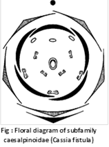
Flower : Bracteate or ebracteate, pedicellate, hermaphrodite, complete, zygomorphic, hypogynous.
Calyx : Sepals 5, polysepalous, imbricate aestivation.
Corolla : Petals 5, polypetalous, ascending imbricate aestivation.
Androecium : 10 stamens, or staminodes are found as in Cassia, free filaments of unequal size, anther lobes bilocular, introrse, versatile.
Gynoecium : Monocarpellary, unilocular, ovary superior, marginal placentation, stigma capitate.
Fruit : Legume.
Floral formula : \[%\,\,\,\,\,\,{{K}_{5}}\,{{C}_{5}}\,{{A}_{1+2+2+2+3\,\left( \text{staminodes} \right)}}\,\text{or}{{\,}_{7+3\,\left( \text{staminodes} \right)}}\,{{G}_{1}}\]
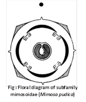
Subfamily – Mimosoideae
Inflorescence : Head or capitulum or spike, flowers arranged in acropetal succession.
Flower : Bracteate or ebracteate, sessile, hermaphrodite, complete actinomorphic, hypogynous, pentamerous.
Calyx : 5 sepals (4 in Mimosa) gamosepalous, connate at the base, valvate aestivation, rarely imbricate (e.g., Parkia).
Corolla : 5 petals (4 in Mimosa) gamopetalous or polypetalous, membranous, valvate aestivation.
Androecium : In most of the members, stamens are indefinite and polyandrous. However, there are only 4 stamens in Mimosa pudica and 10 each in Prosopis and Dichrostachys. Filaments are long, usually connate at the base, sometimes they are coloured and gland dotted. Anthers are dithecous and introrse.
Gynoecium : Monocarpellary, unilocular, ovary superior, style long, cylindrical, stigma single and capitate, marginal placentation.
Fruit : Lomentum.
Floral formula : \[\text{Br}\,\text{or}\,\text{Ebr}\,\,\oplus \,\,\,\,{{K}_{\left( 5 \right)}}\,{{C}_{\left( 5 \right)}}\,\,\text{or}\,{{\,}_{5}}\,{{A}_{\infty }}\,{{G}_{1}}\]
Systematic position
Division : Angiospermae
Class : Monocotyledonae
Series : Coronarieae
Order : Liliales
Family : Liliaceae
Habit : Usually perennial herbs growing by means of rhizomes (e.g., Aloe, Polygonatum), bulbs (e.g., Lilium, Allium) and corms (e.g., Colchicum). Some herbs are annual (e.g., Asphodelus). Shrubs occur in Aloe, Agave, Yucca (Dagger plants, Adam’s Needle), Dracaena (Dragon plant), and Ruscus (Butcher’s Broom). They mostly grow in arid areas and are hence xerophytic (e.g., Aloe, Yucca). Xanthorrhoea of Australia is tree-like. Climbers are seen in Smilax, Gloriosa and species of Asparagus.
Root : Adventitious, fibrous or tuberous (e.g., Asparagus).
Stem : Erect or climbing as Smilex, branched or unbranched, herbaceous, phylloclade as Ruscus. Cladode as Asparagus, Bulb as Allium cepa.
Leaves : Radical or cauline and ramal, cauline and ramal show various types of phyllotaxy (alternate, opposite or whorled), exstipulate, stipulate in Smilax where the stipules are prolonged into tendrils, sessile or petiolate with sheathing leaf bases, venation parallel but reticulate in Smilax, leaves may be scaly, leathery, fleshy or modified into spines (e.g., Asparagus), leaf apex is tendrillar in Gloriosa. The leaves of Phormium tenax (New Zealand Hemp) are 3 metres long and 10 cm broad.
Inflorescence : Recemose, sometimes solitary (e.g., Tulipa, Gloriosa) or umbellate condensed cymes (umbel cyme), e.g., Onion. In several cases the inflorescence possesses a leafless peduncle called scape.
Flower : Bracteate or ebracteate, pedicellate, regular, actinomorphic, zygomorphic in a few cases (e.g., Gilliesia), complete or incomplete, perfect, unisexual in Smilax and Ruscus, hypogynous, generally pentacyclic, trimerous (rarely bimerous or tetramerous). more...
 (b) Progesterone: It is also a steriod hormone secreted by yellow-coloured endocrine gland, called corpus luteum, formed from empty Graafian folicle during more...
 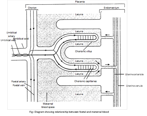 The placenta is fully formed by the end of the third month and it lasts throughout pregnancy. When complete, it is a reddish – brown disc. In the placenta, the foetal blood comes very close to the maternal blood, and this permits the exchange of materials between the two. Food (glucose, amino acids, simple proteins, lipids), water, mineral salts, vitamins, hormones, antibodies and oxygen pass from the maternal blood into the foetal blood, and foetal metabolic wastes, such as carbon dioxide and urea, also water and hormones, pass into the maternal blood. The placenta, thus, serves as the nutritive, respiratory and excretory organ of the foetus. The continuous uptake of oxygen by foetal blood is ensured by the difference in affinity for oxygen between foetal and maternal haemoglobin.
The maternal and foetal blood are not in direct contact in the placenta, because (i) the two may be incompatible; (ii) the pressure of maternal blood is far too high for the foetal blood vessels; and (iii) there must be a check on the passage of harmful materials (blood proteins, germs) into the foetal blood.
(iii) Functions
(1) Placenta helps in the nutrition of the embryo as the nutrients like amino acids, monosugars, vitamins, etc. pass from the maternal blood into foetal blood through placenta.
(2) It also helps in respiration of the embryo as \[{{O}_{2}}\] of the maternal blood more...
The placenta is fully formed by the end of the third month and it lasts throughout pregnancy. When complete, it is a reddish – brown disc. In the placenta, the foetal blood comes very close to the maternal blood, and this permits the exchange of materials between the two. Food (glucose, amino acids, simple proteins, lipids), water, mineral salts, vitamins, hormones, antibodies and oxygen pass from the maternal blood into the foetal blood, and foetal metabolic wastes, such as carbon dioxide and urea, also water and hormones, pass into the maternal blood. The placenta, thus, serves as the nutritive, respiratory and excretory organ of the foetus. The continuous uptake of oxygen by foetal blood is ensured by the difference in affinity for oxygen between foetal and maternal haemoglobin.
The maternal and foetal blood are not in direct contact in the placenta, because (i) the two may be incompatible; (ii) the pressure of maternal blood is far too high for the foetal blood vessels; and (iii) there must be a check on the passage of harmful materials (blood proteins, germs) into the foetal blood.
(iii) Functions
(1) Placenta helps in the nutrition of the embryo as the nutrients like amino acids, monosugars, vitamins, etc. pass from the maternal blood into foetal blood through placenta.
(2) It also helps in respiration of the embryo as \[{{O}_{2}}\] of the maternal blood more... 

 Flower : Bracteate or ebracteate, pedicellate, hermaphrodite, complete, zygomorphic, hypogynous.
Calyx : Sepals 5, polysepalous, imbricate aestivation.
Corolla : Petals 5, polypetalous, ascending imbricate aestivation.
Androecium : 10 stamens, or staminodes are found as in Cassia, free filaments of unequal size, anther lobes bilocular, introrse, versatile.
Gynoecium : Monocarpellary, unilocular, ovary superior, marginal placentation, stigma capitate.
Fruit : Legume.
Floral formula : \[%\,\,\,\,\,\,{{K}_{5}}\,{{C}_{5}}\,{{A}_{1+2+2+2+3\,\left( \text{staminodes} \right)}}\,\text{or}{{\,}_{7+3\,\left( \text{staminodes} \right)}}\,{{G}_{1}}\]
Subfamily – Mimosoideae
Inflorescence : Head or capitulum or spike, flowers arranged in acropetal succession.
Flower : Bracteate or ebracteate, sessile, hermaphrodite, complete actinomorphic, hypogynous, pentamerous.
Calyx : 5 sepals (4 in Mimosa) gamosepalous, connate at the base, valvate aestivation, rarely imbricate (e.g., Parkia).
Corolla : 5 petals (4 in Mimosa) gamopetalous or polypetalous, membranous, valvate aestivation.
Androecium : In most of the members, stamens are indefinite and polyandrous. However, there are only 4 stamens in Mimosa pudica and 10 each in Prosopis and Dichrostachys. Filaments are long, usually connate at the base, sometimes they are coloured and gland dotted. Anthers are dithecous and introrse.
Gynoecium : Monocarpellary, unilocular, ovary superior, style long, cylindrical, stigma single and capitate, marginal placentation.
Fruit : Lomentum.
Floral formula : \[\text{Br}\,\text{or}\,\text{Ebr}\,\,\oplus \,\,\,\,{{K}_{\left( 5 \right)}}\,{{C}_{\left( 5 \right)}}\,\,\text{or}\,{{\,}_{5}}\,{{A}_{\infty }}\,{{G}_{1}}\]
Flower : Bracteate or ebracteate, pedicellate, hermaphrodite, complete, zygomorphic, hypogynous.
Calyx : Sepals 5, polysepalous, imbricate aestivation.
Corolla : Petals 5, polypetalous, ascending imbricate aestivation.
Androecium : 10 stamens, or staminodes are found as in Cassia, free filaments of unequal size, anther lobes bilocular, introrse, versatile.
Gynoecium : Monocarpellary, unilocular, ovary superior, marginal placentation, stigma capitate.
Fruit : Legume.
Floral formula : \[%\,\,\,\,\,\,{{K}_{5}}\,{{C}_{5}}\,{{A}_{1+2+2+2+3\,\left( \text{staminodes} \right)}}\,\text{or}{{\,}_{7+3\,\left( \text{staminodes} \right)}}\,{{G}_{1}}\]
Subfamily – Mimosoideae
Inflorescence : Head or capitulum or spike, flowers arranged in acropetal succession.
Flower : Bracteate or ebracteate, sessile, hermaphrodite, complete actinomorphic, hypogynous, pentamerous.
Calyx : 5 sepals (4 in Mimosa) gamosepalous, connate at the base, valvate aestivation, rarely imbricate (e.g., Parkia).
Corolla : 5 petals (4 in Mimosa) gamopetalous or polypetalous, membranous, valvate aestivation.
Androecium : In most of the members, stamens are indefinite and polyandrous. However, there are only 4 stamens in Mimosa pudica and 10 each in Prosopis and Dichrostachys. Filaments are long, usually connate at the base, sometimes they are coloured and gland dotted. Anthers are dithecous and introrse.
Gynoecium : Monocarpellary, unilocular, ovary superior, style long, cylindrical, stigma single and capitate, marginal placentation.
Fruit : Lomentum.
Floral formula : \[\text{Br}\,\text{or}\,\text{Ebr}\,\,\oplus \,\,\,\,{{K}_{\left( 5 \right)}}\,{{C}_{\left( 5 \right)}}\,\,\text{or}\,{{\,}_{5}}\,{{A}_{\infty }}\,{{G}_{1}}\]
 Systematic position
Division : Angiospermae
Class : Monocotyledonae
Series : Coronarieae
Order : Liliales
Family : Liliaceae
Habit : Usually perennial herbs growing by means of rhizomes (e.g., Aloe, Polygonatum), bulbs (e.g., Lilium, Allium) and corms (e.g., Colchicum). Some herbs are annual (e.g., Asphodelus). Shrubs occur in Aloe, Agave, Yucca (Dagger plants, Adam’s Needle), Dracaena (Dragon plant), and Ruscus (Butcher’s Broom). They mostly grow in arid areas and are hence xerophytic (e.g., Aloe, Yucca). Xanthorrhoea of Australia is tree-like. Climbers are seen in Smilax, Gloriosa and species of Asparagus.
Root : Adventitious, fibrous or tuberous (e.g., Asparagus).
Stem : Erect or climbing as Smilex, branched or unbranched, herbaceous, phylloclade as Ruscus. Cladode as Asparagus, Bulb as Allium cepa.
Leaves : Radical or cauline and ramal, cauline and ramal show various types of phyllotaxy (alternate, opposite or whorled), exstipulate, stipulate in Smilax where the stipules are prolonged into tendrils, sessile or petiolate with sheathing leaf bases, venation parallel but reticulate in Smilax, leaves may be scaly, leathery, fleshy or modified into spines (e.g., Asparagus), leaf apex is tendrillar in Gloriosa. The leaves of Phormium tenax (New Zealand Hemp) are 3 metres long and 10 cm broad.
Inflorescence : Recemose, sometimes solitary (e.g., Tulipa, Gloriosa) or umbellate condensed cymes (umbel cyme), e.g., Onion. In several cases the inflorescence possesses a leafless peduncle called scape.
Flower : Bracteate or ebracteate, pedicellate, regular, actinomorphic, zygomorphic in a few cases (e.g., Gilliesia), complete or incomplete, perfect, unisexual in Smilax and Ruscus, hypogynous, generally pentacyclic, trimerous (rarely bimerous or tetramerous).
Systematic position
Division : Angiospermae
Class : Monocotyledonae
Series : Coronarieae
Order : Liliales
Family : Liliaceae
Habit : Usually perennial herbs growing by means of rhizomes (e.g., Aloe, Polygonatum), bulbs (e.g., Lilium, Allium) and corms (e.g., Colchicum). Some herbs are annual (e.g., Asphodelus). Shrubs occur in Aloe, Agave, Yucca (Dagger plants, Adam’s Needle), Dracaena (Dragon plant), and Ruscus (Butcher’s Broom). They mostly grow in arid areas and are hence xerophytic (e.g., Aloe, Yucca). Xanthorrhoea of Australia is tree-like. Climbers are seen in Smilax, Gloriosa and species of Asparagus.
Root : Adventitious, fibrous or tuberous (e.g., Asparagus).
Stem : Erect or climbing as Smilex, branched or unbranched, herbaceous, phylloclade as Ruscus. Cladode as Asparagus, Bulb as Allium cepa.
Leaves : Radical or cauline and ramal, cauline and ramal show various types of phyllotaxy (alternate, opposite or whorled), exstipulate, stipulate in Smilax where the stipules are prolonged into tendrils, sessile or petiolate with sheathing leaf bases, venation parallel but reticulate in Smilax, leaves may be scaly, leathery, fleshy or modified into spines (e.g., Asparagus), leaf apex is tendrillar in Gloriosa. The leaves of Phormium tenax (New Zealand Hemp) are 3 metres long and 10 cm broad.
Inflorescence : Recemose, sometimes solitary (e.g., Tulipa, Gloriosa) or umbellate condensed cymes (umbel cyme), e.g., Onion. In several cases the inflorescence possesses a leafless peduncle called scape.
Flower : Bracteate or ebracteate, pedicellate, regular, actinomorphic, zygomorphic in a few cases (e.g., Gilliesia), complete or incomplete, perfect, unisexual in Smilax and Ruscus, hypogynous, generally pentacyclic, trimerous (rarely bimerous or tetramerous).


