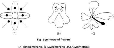
 (4) Arrangement of floral organs : On the basis of arrangement of floral organs, three types of flowers more...
 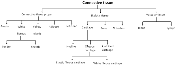 Connective tissue proper
 possess soft viscous semisolid or semi-fluid matrix.
Connective tissue proper
Connective tissue proper possess soft viscous semisolid or semi-fluid matrix.
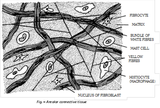 (i) Areolar Tissue: Areolar tissue is loose connective tissue, possess transparent gelatinous, highly vascular and sticky matrix which have variety of cells and fibres. It allows movement of part connected by it (Muscle and their compound). Areolar tissue mainly consist more...
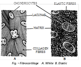
 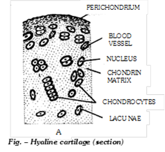 (2) Fibro cartilage (White fibrous cartilage): In this cartilage, the small amount of matrix of cartilage is packed with large number of bundles of thick white (collagen) fibres. So it is toughest and less flexible. Between the bundles of white fibres, there are scattered lacunae, each containing a chondrocyte. It is found in intervertebral discs and acts as shock absorber. It is also found in pubic symphysis and helps in parturition (child birth). The intervertebral discs remain contracted when the body is active, but relaxed when the body is at rest. That is why, our body becomes a bit taller during sleep and after death.
 (3) Elastic more...
 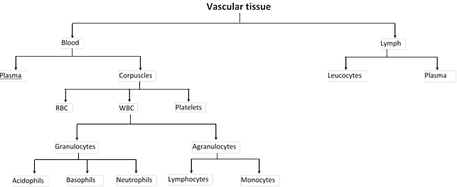 (i) Blood: In chordates, and in annelids amongst the non chordates, the blood is a red and opaque fluid of salty taste and peculiar smell. It is a little heavier than water. Its specific gravity and viscosity is 1.04 - 1.07 and 4.7 respectively. Its is 7.4 so it is slightly alkaline. In human beings, the quantity of blood is about 7% to 8% of total body weight. Thus a person, weighing about 70 kg has about 5 to 6 litres of blood, occupying about 1/13th part of the body by volume. Percentage of blood in women is slightly lower. The study of blood is called haematology. It is red coloured liquid connective tissue which originates from the mesoderm. It reaches into the various organs through the blood vessels and transports various chemical substances between different tissues. During embryonic state, the blood is mainly formed in the liver but little blood is also formed in the spleen and ribs. In adults, the blood is formed in the red bone marrow. The blood formation is called as haemopoiesis.
(ii) Plasma: It constitutes about 5% of body weight. It represents matrix of blood. Plasma is slightly alkaline and transparent. It forms 55-60% by volume of blood. Plasma contains: Water (91-92%), Solid (8-9%). Plasma solid part consists of organic (7%) and inorganic (1%) substances which are as follows:
(a) Organic constituents of plasma: Some are its own constituents, while others are those which are transported by it. All these are divisible into following categories:
(1) Plasma proteins: Protein constitute about 7% part of plasma and remain in it as colloid particles. These mainly include albumins, globulins, prothrombin and fibrinogen.
(i) Blood: In chordates, and in annelids amongst the non chordates, the blood is a red and opaque fluid of salty taste and peculiar smell. It is a little heavier than water. Its specific gravity and viscosity is 1.04 - 1.07 and 4.7 respectively. Its is 7.4 so it is slightly alkaline. In human beings, the quantity of blood is about 7% to 8% of total body weight. Thus a person, weighing about 70 kg has about 5 to 6 litres of blood, occupying about 1/13th part of the body by volume. Percentage of blood in women is slightly lower. The study of blood is called haematology. It is red coloured liquid connective tissue which originates from the mesoderm. It reaches into the various organs through the blood vessels and transports various chemical substances between different tissues. During embryonic state, the blood is mainly formed in the liver but little blood is also formed in the spleen and ribs. In adults, the blood is formed in the red bone marrow. The blood formation is called as haemopoiesis.
(ii) Plasma: It constitutes about 5% of body weight. It represents matrix of blood. Plasma is slightly alkaline and transparent. It forms 55-60% by volume of blood. Plasma contains: Water (91-92%), Solid (8-9%). Plasma solid part consists of organic (7%) and inorganic (1%) substances which are as follows:
(a) Organic constituents of plasma: Some are its own constituents, while others are those which are transported by it. All these are divisible into following categories:
(1) Plasma proteins: Protein constitute about 7% part of plasma and remain in it as colloid particles. These mainly include albumins, globulins, prothrombin and fibrinogen.
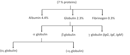 Globulins are mainly formed by plasma cells in lymphoid organs. Other plasma proteins are mainly formed in liver. These render the plasma viscous, and maintain its osmotic pressure (7.5 atmospheric) and pH. Prothrombin and Fibrinogen are essential for blood clotting. Albumins are mainly more...
 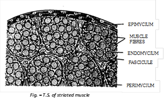 (a) Structure of striated muscles:
Each striated muscle consists of numerous muscle fibres segregated into several small and parallel bundles, called Fasciculi. Fibres of each fascicule are bound together by a connective tissue sheath, called endomycium. All fasciculi of a muscle are bound together by a connective tissue termed perimycium which also forms a sheath around each fascicule. Similarly, the whole muscle itself is covered by a connective tissue sheath, called epimycium. The latter extends as a tendons at each end of the muscle to insert it on to bones. Endomycium, more...
(a) Structure of striated muscles:
Each striated muscle consists of numerous muscle fibres segregated into several small and parallel bundles, called Fasciculi. Fibres of each fascicule are bound together by a connective tissue sheath, called endomycium. All fasciculi of a muscle are bound together by a connective tissue termed perimycium which also forms a sheath around each fascicule. Similarly, the whole muscle itself is covered by a connective tissue sheath, called epimycium. The latter extends as a tendons at each end of the muscle to insert it on to bones. Endomycium, more... 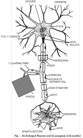 (a) Structure of neurons: A neuron is a nerve cell with all its branches. Neuron is formed from neuroblast. It is the structural and functional unit of nervous system. It is the longest cell of the body.
(1) Cyton: It is also called perikaryon or soma or cell body. Its granular cytoplasm is called neuroplasm which has following structures :
(i) A large, spherical, centrally placed nucleus with a single nucleolus.
(ii) Numerous fine threads called neurofibrils for the conduction of nerve impulses.
(iii) A number of small, basophilic granules called Nissl’s granules formed of rough endoplasmic reticulum with ribosomes and are sites of protein synthesis.
(iv) Neuroplasm has large number of mitochondria to provide high energy for impulse conduction.
(v) Neuroplasm may have melanophores with melanin pigment and lipochromes with orange or yellow pigment.
(vi) A mature neuron has no centriole, so it cannot divide.
(vii) A “Barr body” is often seen abutting against the inner surface of nuclear membrane of cytons in females. This has been proved to be a transformed ‘X’ chromosome.
(viii) Certain neurons having flask-shaped cytons and called purkinje cells, occur in the cerebellum of the brain.
(2) Neuron processes: The processes of neurons, called neurites, extend varying distances from the cyton and are of two types - dendrites or dendrons and an axon or axis cylinder (neuraxon).
(i) more...
(a) Structure of neurons: A neuron is a nerve cell with all its branches. Neuron is formed from neuroblast. It is the structural and functional unit of nervous system. It is the longest cell of the body.
(1) Cyton: It is also called perikaryon or soma or cell body. Its granular cytoplasm is called neuroplasm which has following structures :
(i) A large, spherical, centrally placed nucleus with a single nucleolus.
(ii) Numerous fine threads called neurofibrils for the conduction of nerve impulses.
(iii) A number of small, basophilic granules called Nissl’s granules formed of rough endoplasmic reticulum with ribosomes and are sites of protein synthesis.
(iv) Neuroplasm has large number of mitochondria to provide high energy for impulse conduction.
(v) Neuroplasm may have melanophores with melanin pigment and lipochromes with orange or yellow pigment.
(vi) A mature neuron has no centriole, so it cannot divide.
(vii) A “Barr body” is often seen abutting against the inner surface of nuclear membrane of cytons in females. This has been proved to be a transformed ‘X’ chromosome.
(viii) Certain neurons having flask-shaped cytons and called purkinje cells, occur in the cerebellum of the brain.
(2) Neuron processes: The processes of neurons, called neurites, extend varying distances from the cyton and are of two types - dendrites or dendrons and an axon or axis cylinder (neuraxon).
(i) more... 


You need to login to perform this action.
You will be redirected in
3 sec

