
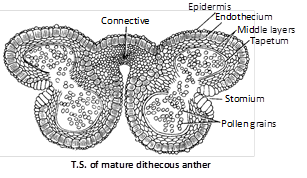
 The pollen sacs are surrounded by following 4 layers :
(i) Epidermis : This is the outermost single layered and protective. In Arceuthobium, cells of epidermis develops a fibrous thickening and the epidermis is designated as exothecium.
(ii) Endothecium : Inner to epidermis, there is a single layer of radially elongated cells. Cells of endothecium develop fibrous thickening (made up of cellulose with a little pectin and lignin) which help in the dehiscence of anther. In between these cells, a few cells without thickening are also present. These thin walled cells collectively form the stomium.
(iii) Middle layer : Three to four layers of thin walled cells situated just below the endothecium are known as middle layers. Cells of this layer are ephemeral and degenerate to provide nourishment to growing microspore mother cells.
(iv) Tapetum : This is the innermost layer of the wall. The cells are multinucleate(undergo endopolyploidy) and polyploid. Tapetal cells are nutritive.
In these cells the Ubisch bodies are present which help in the ornamentation of microspore walls. A compound sporopollenin is secreted in the exine of microspore wall. According to Periasamy and Swamy (1966), developmentally the tapetum has dual nature.
The tapetum is of two types
(a) Amoeboid or Periplasmodial tapetum.
(b) Secretory or Glandular tapetum.
(2) Development of anther and formation of microspores (Pollen grains) : The young anther consists of homogenous mass of paranchymatous cells surrounded by epidermis. It soon becomes four lobed. In each of the four lobes, some of the hypodermal cells begin to act as archesporial initials. Each archesporial initial divides into an outer primary parietal cell and an inner primary sporogenous cell. The primary parietal cell divides to form 3-5 wall layers, i.e., endothecium, middle layers and tapetum. The primary sporogenous cells divide to produce a mass of sporogenous cells or microsporocytes.
Each microspore mother cell divides meiotically to form four haploid microspores or pollen grains and remains arranged in tetrads. The arrangement in the tetrads can be tetrahedral, isobilateral, linear, T-shaped and decussate.
Now the microspores are separated from tetrad. In Drosera, Typha, Elodea, Hydrilla, etc. all the four pollen grains do not separate and thus form compound pollen grains. In the members of the family Cyperaceae (Cyprus), out of 4 pollen in a tetrad, 3 degenerate and one remains alive. So one meiosis produces one pollen. Sometimes more than four pollens are produced from one microspore mother more...
The pollen sacs are surrounded by following 4 layers :
(i) Epidermis : This is the outermost single layered and protective. In Arceuthobium, cells of epidermis develops a fibrous thickening and the epidermis is designated as exothecium.
(ii) Endothecium : Inner to epidermis, there is a single layer of radially elongated cells. Cells of endothecium develop fibrous thickening (made up of cellulose with a little pectin and lignin) which help in the dehiscence of anther. In between these cells, a few cells without thickening are also present. These thin walled cells collectively form the stomium.
(iii) Middle layer : Three to four layers of thin walled cells situated just below the endothecium are known as middle layers. Cells of this layer are ephemeral and degenerate to provide nourishment to growing microspore mother cells.
(iv) Tapetum : This is the innermost layer of the wall. The cells are multinucleate(undergo endopolyploidy) and polyploid. Tapetal cells are nutritive.
In these cells the Ubisch bodies are present which help in the ornamentation of microspore walls. A compound sporopollenin is secreted in the exine of microspore wall. According to Periasamy and Swamy (1966), developmentally the tapetum has dual nature.
The tapetum is of two types
(a) Amoeboid or Periplasmodial tapetum.
(b) Secretory or Glandular tapetum.
(2) Development of anther and formation of microspores (Pollen grains) : The young anther consists of homogenous mass of paranchymatous cells surrounded by epidermis. It soon becomes four lobed. In each of the four lobes, some of the hypodermal cells begin to act as archesporial initials. Each archesporial initial divides into an outer primary parietal cell and an inner primary sporogenous cell. The primary parietal cell divides to form 3-5 wall layers, i.e., endothecium, middle layers and tapetum. The primary sporogenous cells divide to produce a mass of sporogenous cells or microsporocytes.
Each microspore mother cell divides meiotically to form four haploid microspores or pollen grains and remains arranged in tetrads. The arrangement in the tetrads can be tetrahedral, isobilateral, linear, T-shaped and decussate.
Now the microspores are separated from tetrad. In Drosera, Typha, Elodea, Hydrilla, etc. all the four pollen grains do not separate and thus form compound pollen grains. In the members of the family Cyperaceae (Cyprus), out of 4 pollen in a tetrad, 3 degenerate and one remains alive. So one meiosis produces one pollen. Sometimes more than four pollens are produced from one microspore mother more... 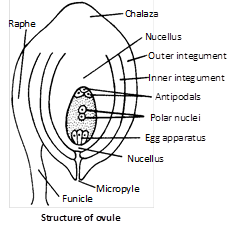 The residual part of nucellus in the mature seed is called perisperm. In the centre of the nucellus is situated a female gametophyte known as embryo sac.
Following are the conditions seen in ovule in relation to integuments :
(i) Unitegmic : Ovule with a single integument, e.g., sympetalous or gamopetalous dicotyledons.
(ii) Bitegmic : Ovule with two integuments as in polypetalous (Archichlamydeae) dicotyledons and monocotyledons.
(iii) Aril : This is a collar-like outgrowth from the base of the ovule and forms third integument. Aril is found in litchi, nutmeg, etc.
(iv) Caruncle : It is formed as an outgrowth of the outer integument in the micropylar region. Caruncle is common in the ovules of Euphorbiaceae. e.g., Castor (Ricinus).
(v) Ategmic : In some parasites like Loranthus, Viscum, Santalum etc., there is no integument. Such an ovule is called ategmic.
(2) Kinds of ovules : Depending upon the shape and orientation, the ovules of angiosperms are classified into following types :
The residual part of nucellus in the mature seed is called perisperm. In the centre of the nucellus is situated a female gametophyte known as embryo sac.
Following are the conditions seen in ovule in relation to integuments :
(i) Unitegmic : Ovule with a single integument, e.g., sympetalous or gamopetalous dicotyledons.
(ii) Bitegmic : Ovule with two integuments as in polypetalous (Archichlamydeae) dicotyledons and monocotyledons.
(iii) Aril : This is a collar-like outgrowth from the base of the ovule and forms third integument. Aril is found in litchi, nutmeg, etc.
(iv) Caruncle : It is formed as an outgrowth of the outer integument in the micropylar region. Caruncle is common in the ovules of Euphorbiaceae. e.g., Castor (Ricinus).
(v) Ategmic : In some parasites like Loranthus, Viscum, Santalum etc., there is no integument. Such an ovule is called ategmic.
(2) Kinds of ovules : Depending upon the shape and orientation, the ovules of angiosperms are classified into following types :
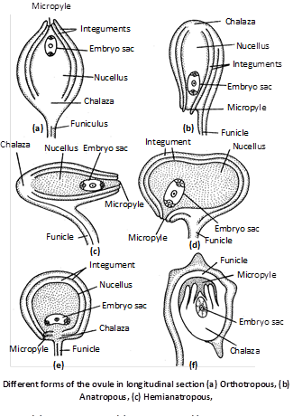 (i) Orthotropous or Atropus : The micropyle, chalaza and funicle are in straight line. This is most primitive type of ovules. e.g., Betel, Piper, Polygonum.
(ii) Anatropous : The body of the ovule is completely inverted (turn at 180o angle ) so that micropyle and hilum come to lie very close to each other. e.g., 82% of angiosperm families.
(iii) Hemianatropous : Ovule turns at 90o angle upon the funicle or body of ovule is at right angle to the funicle e.g., Ranunculus.
(iv) Campylotropous : Ovule is circled more or less at right angle to funicle. Micropylar end is bent down slightly. e.g., in members of Leguminosae and Cruciferae.
(v) Amphitropous : Curvature of ovule is more and embryo sac becomes curved like horse shoe e.g. Lemna, Poppy, Alisma.
(vi) Circinotropous : The ovule is initially more...
(i) Orthotropous or Atropus : The micropyle, chalaza and funicle are in straight line. This is most primitive type of ovules. e.g., Betel, Piper, Polygonum.
(ii) Anatropous : The body of the ovule is completely inverted (turn at 180o angle ) so that micropyle and hilum come to lie very close to each other. e.g., 82% of angiosperm families.
(iii) Hemianatropous : Ovule turns at 90o angle upon the funicle or body of ovule is at right angle to the funicle e.g., Ranunculus.
(iv) Campylotropous : Ovule is circled more or less at right angle to funicle. Micropylar end is bent down slightly. e.g., in members of Leguminosae and Cruciferae.
(v) Amphitropous : Curvature of ovule is more and embryo sac becomes curved like horse shoe e.g. Lemna, Poppy, Alisma.
(vi) Circinotropous : The ovule is initially more... 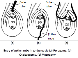 (3) Entry of pollen tube into embryo sac : The pollen tube enters the embryo sac only from the micropylar end irrespective of its mode of entry into the ovule. The pollen tube either passes between a synergid and the egg cell or enters into one of the synergids through filiform apparatus. The synergids direct the growth of pollen tube by secreting some chemical substances (chemotropic secretion). The tip of pollen tube enters into one synergid. The penetrated synergid starts degenerating. After penetration, the tip of pollen tube enlarge and ruptures releasing most of its contents including the two male gametes and the vegetative nucleus into the synergid.
(4) Double fertilization : The nuclei of both the male gametes are released in the embryo sac. One male gamete fuses with the egg to form the diploid zygote. The process is called syngamy or generative fertilization. This syngamy was discovered by Strasburger (1884).
The diploid zygote finally develops into embryo. The other male gamete fuses with the two polar nuclei (or secondary nucleus) to form the triploid primary endosperm nucleus. The process is called triple fusion or vegetative fertilization. These two acts of fertilizations constitute the process of double fertilization. The process was discovered by S.G. Nawaschin (1898) and Guignard in Lilium and Frittillaria. Double fertilization occurs in angiosperms more...
(3) Entry of pollen tube into embryo sac : The pollen tube enters the embryo sac only from the micropylar end irrespective of its mode of entry into the ovule. The pollen tube either passes between a synergid and the egg cell or enters into one of the synergids through filiform apparatus. The synergids direct the growth of pollen tube by secreting some chemical substances (chemotropic secretion). The tip of pollen tube enters into one synergid. The penetrated synergid starts degenerating. After penetration, the tip of pollen tube enlarge and ruptures releasing most of its contents including the two male gametes and the vegetative nucleus into the synergid.
(4) Double fertilization : The nuclei of both the male gametes are released in the embryo sac. One male gamete fuses with the egg to form the diploid zygote. The process is called syngamy or generative fertilization. This syngamy was discovered by Strasburger (1884).
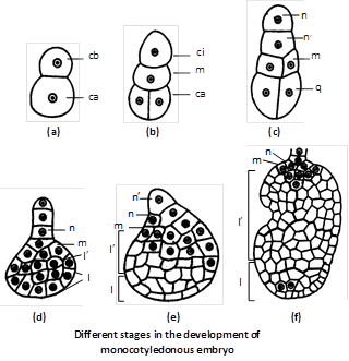
The diploid zygote finally develops into embryo. The other male gamete fuses with the two polar nuclei (or secondary nucleus) to form the triploid primary endosperm nucleus. The process is called triple fusion or vegetative fertilization. These two acts of fertilizations constitute the process of double fertilization. The process was discovered by S.G. Nawaschin (1898) and Guignard in Lilium and Frittillaria. Double fertilization occurs in angiosperms more... 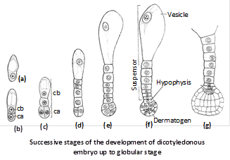 (ii) In monocotyledons : The normal type of monocot embryo development has been studied in Sagittaria sagittaefolia. The early development of dicot and monocot embryos is similar upto globular stage. Later on differentiation starts. Suspensor is single celled and vascular. There is only one terminal cotyledon called scutellum (shield shaped). In grasses the second cotyledon is reduced called epiblast.
The basal cell (cb) divides by a transverse wall into two cells – ci and m. The cell ci divides once again to form n and n’ cells. Of these n’ is the outermost which develops into suspensor. The cell n forms parts of root cap the cell m contributes to the remaining part of root cap and a part of the radicle.
The terminal cell (ca) divides by two vertical walls, at right angles to one another. This results in the formation of a quadrant (q). Cells of the quadrant divide periclinally differentiating into the peripheral cells and the inner group of cells. The repeated divisions in both peripheral and central group of cells results in the formation of two regions –l and l’. Region l produces the lower part of cotyledon while upper part of cotyledon, hypocotyl and plumule are formed by l’ region.
(ii) In monocotyledons : The normal type of monocot embryo development has been studied in Sagittaria sagittaefolia. The early development of dicot and monocot embryos is similar upto globular stage. Later on differentiation starts. Suspensor is single celled and vascular. There is only one terminal cotyledon called scutellum (shield shaped). In grasses the second cotyledon is reduced called epiblast.
The basal cell (cb) divides by a transverse wall into two cells – ci and m. The cell ci divides once again to form n and n’ cells. Of these n’ is the outermost which develops into suspensor. The cell n forms parts of root cap the cell m contributes to the remaining part of root cap and a part of the radicle.
The terminal cell (ca) divides by two vertical walls, at right angles to one another. This results in the formation of a quadrant (q). Cells of the quadrant divide periclinally differentiating into the peripheral cells and the inner group of cells. The repeated divisions in both peripheral and central group of cells results in the formation of two regions –l and l’. Region l produces the lower part of cotyledon while upper part of cotyledon, hypocotyl and plumule are formed by l’ region.
 (2) Polyembryony : Occurrence of more than two embryo in the seed is known as polyembryony. It was discovered by A.V. Leeuwenhock (1719) in Citrus. It may be :
(i) Cleavage polyembryony : Due to cleavage of zygote or proembryo into two or more embryos and each split part develops into more...
(2) Polyembryony : Occurrence of more than two embryo in the seed is known as polyembryony. It was discovered by A.V. Leeuwenhock (1719) in Citrus. It may be :
(i) Cleavage polyembryony : Due to cleavage of zygote or proembryo into two or more embryos and each split part develops into more... 





You need to login to perform this action.
You will be redirected in
3 sec