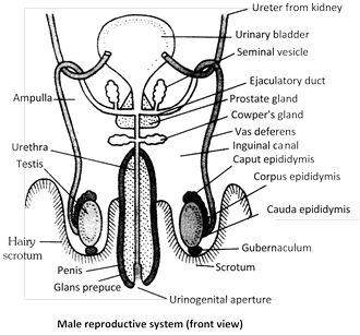
 A spermatic cord connects testis with abdominal cavity. It consists of connective tissue that encloses an artery, a vein, a lymph vessel, a nerve, cremaster muscle and a vas deferens. A testis rests in it chamber over pad called gubernaculum.
The scrotal sac of male homologous to female's labia majora.
A spermatic cord connects testis with abdominal cavity. It consists of connective tissue that encloses an artery, a vein, a lymph vessel, a nerve, cremaster muscle and a vas deferens. A testis rests in it chamber over pad called gubernaculum.
The scrotal sac of male homologous to female's labia majora.
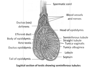 (2) Human Testes : The testes are the primary sex organs. They are about \[45\,\,cm\] long, 2.5 cm wide and 3 cm thick. They are suspended in the scrotal sacs by spermatic cords. Each testes weights about \[10-15\text{ }gms.\]
Each testis has three coverings - tunica vaginalis, tunica albuginea and tunica vasculosa.
In growth of the tunica albuginea, called septa, divide the testis into some 200 to 300 lobules. Each testicular lobule contains \[13\] highly convoluted seminiferous tubules, blood vessels and nerve embedded in loose connective tissue. A total of about 750 seminiferous tubules occur in each testis.
(2) Human Testes : The testes are the primary sex organs. They are about \[45\,\,cm\] long, 2.5 cm wide and 3 cm thick. They are suspended in the scrotal sacs by spermatic cords. Each testes weights about \[10-15\text{ }gms.\]
Each testis has three coverings - tunica vaginalis, tunica albuginea and tunica vasculosa.
In growth of the tunica albuginea, called septa, divide the testis into some 200 to 300 lobules. Each testicular lobule contains \[13\] highly convoluted seminiferous tubules, blood vessels and nerve embedded in loose connective tissue. A total of about 750 seminiferous tubules occur in each testis.
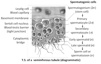 Each seminiferous tubules is lined by germinal epithelium, seminiferous tubules is the site of spermatogenesis. The process occurs in waves along the length of the tubule, taking about 9 weeks (63 days) to complete in man. Seminiferous tubules contain 3 types of cells -
(i) Germ cells : Germ cells or primordial germ cells arise from yolk sac endoderm and enter the testes early in development. These are spermatogenic cells, by mitotic divisions, produce spermatogonia into the lumen of the seminiferous tubule. The spermatogonia grow into primary spermatocytes which undergo meiosis, producing haploid cells, first secondary spermatocytes and then spermatids. Spermatids differentiate by a more...
Each seminiferous tubules is lined by germinal epithelium, seminiferous tubules is the site of spermatogenesis. The process occurs in waves along the length of the tubule, taking about 9 weeks (63 days) to complete in man. Seminiferous tubules contain 3 types of cells -
(i) Germ cells : Germ cells or primordial germ cells arise from yolk sac endoderm and enter the testes early in development. These are spermatogenic cells, by mitotic divisions, produce spermatogonia into the lumen of the seminiferous tubule. The spermatogonia grow into primary spermatocytes which undergo meiosis, producing haploid cells, first secondary spermatocytes and then spermatids. Spermatids differentiate by a more... | Time from fertilization | Stage/organs | Event | |||||||||||||||
| 24 hours | Cleavage | Embryo is at two-cell stage | |||||||||||||||
| 3 days | Modula | The module reaches uterus | |||||||||||||||
| 7 days | Blastocyst | Implantation of blastocyst begins | |||||||||||||||
| 2.5 weeks | Notochord | Notochord formed, differentiation of tissues that will give rise to heart, blood cells formed in yolk sac and chorionic. | |||||||||||||||
| 3.5 weeks | Organ system | Neural tube formed, primordial eye and ear vesicle, pharyngeal pouches formed, liver bud differentiates, respiratory system and thyroid gland begin to develop, heart tube bends and begins to beat, blood vessels are formed. | |||||||||||||||
| 4 weeks | Limb buds | Development and appearance of limb buds, brain forms three primary vesicles. | |||||||||||||||
| 2 months | Muscles and gonads | Muscles differentiate, embryo capable of movement, gonads distinguishable as testes or ovaries, ossification of bones begins, cerebral cortex is differentiated, blood vessels assume final position. | |||||||||||||||
| 3 months | Sex differentiation | By external examination sex can be determined, notochord degenerates, lymph glands develop. | |||||||||||||||
| 4 months | Face | Face begins to look human, eye, ear and nose look ‘normal’, cerebral lobes differentiate. | |||||||||||||||
| 6-9 months (third trimester) | Lanugo (hairs) body growth |
Lanugo appear but more...
Embryonic development involves following dynamic changes and identifiable process.
(1) Gametogenesis : It involve the formation of haploid sex cells or gametes called sperms and ova from diploid primary germ cells called gametogonia present in the reproductive organs called gonads (testes and ovary). It is of two types
(i) Spermatogenesis : Formation of sperm.
(ii) Oogenesis : Formation of ova
(2) Fertilization : It involve the fusion of haploid male and female gametes to form diploid zygote. The fusion of gametic pronuclei is called Karyogamy while the mixing of two sets of chromosomes of two gametes is called amphimixis.
(3) Cleavage : It includes the rapid mitotic division of the zygote to form a single layered hollow spherical larva called blastula and its formation is called blastulation.
(4) Implantation : The process of attachment of the blastocyst (mammalian blastula) on the endometrium of the uterus is called implantation.
(5) Gastrulation : It includes the mass and orderly migration of the organ specific areas from the surface of blastula to their predetermined position which finally produces a 3 layered gastrula larva. It is with 3 primary layers.
(6) Organogenesis : It includes the formation of specific organs system from three primary germ layers of gastrula and also includes the morphogenesis and differentiation.
Important Tips
Historical background of Embryonic Development:
(i) Definition: Placenta is defined as a temporary intimate mechanical and physiological connection between foetal and maternal tissues for the nutrition, respiration and excretion of the foetus.
(ii) Structure: Human placenta consist of chorion only. Hence, it is called a chorionic placenta. Allantois remains small. The allantoic blood vessels, however, extend to vascularize it. A large number of branching villi from the vascular chorion penetrate the corresponding pits, the crypts, formed in the uterine wall. The latter becomes very thick and highly vascular to receive the villi. The intimate connection established between the foetal membrane and the uterine wall is known as the placenta. The placenta has two parts: the part contributed by the foetus, i.e., chorionic villi, is called the foetal placenta; and the part shared by the mother, i.e., part of uterine wall, is termed the maternal placenta. The chorionic villi receive blood from the embryo by umbilical artery and return it by umbilical vein. These blood vessels are derived from the allantois and run between the foetus and the uterine wall in the tough umbilical cord covered with cells derived from the amnion and chorion. The choroinic villi come to lie in uterine lacunae that receive blood from the uterine arteriole and return it by uterine venule. The cells forming the wall of chorionic villi bear microvilli which increase their surface area for quick and adequate exchange of materials by diffusion, active transport and pinocytosis.
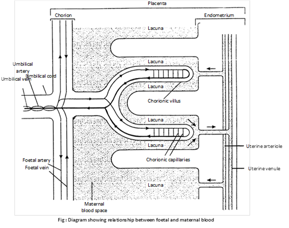 The placenta is fully formed by the end of the third month and it lasts throughout pregnancy. When complete, it is a reddish – brown disc. In the placenta, the foetal blood comes very close to the maternal blood, and this permits the exchange of materials between the two. Food (glucose, amino acids, simple proteins, lipids), water, mineral salts, vitamins, hormones, antibodies and oxygen pass from the maternal blood into the foetal blood, and foetal metabolic wastes, such as carbon dioxide and urea, also water and hormones, pass into the maternal blood. The placenta, thus, serves as the nutritive, respiratory and excretory organ of the foetus. The continuous uptake of oxygen by foetal blood is ensured by the difference in affinity for oxygen between foetal and maternal haemoglobin.
The maternal and foetal blood are not in direct contact in the placenta, because (i) the two may be incompatible; (ii) the pressure of maternal blood is far too high for the foetal blood vessels; and (iii) there must be a check on the passage of harmful materials (blood proteins, germs) into the foetal blood.
(iii) Functions
(1) Placenta helps in the nutrition of the embryo as the nutrients like amino acids, monosugars, vitamins, etc. pass from the maternal blood into foetal blood through placenta.
(2) It also helps in respiration of the embryo as \[{{O}_{2}}\] of the maternal more...
The placenta is fully formed by the end of the third month and it lasts throughout pregnancy. When complete, it is a reddish – brown disc. In the placenta, the foetal blood comes very close to the maternal blood, and this permits the exchange of materials between the two. Food (glucose, amino acids, simple proteins, lipids), water, mineral salts, vitamins, hormones, antibodies and oxygen pass from the maternal blood into the foetal blood, and foetal metabolic wastes, such as carbon dioxide and urea, also water and hormones, pass into the maternal blood. The placenta, thus, serves as the nutritive, respiratory and excretory organ of the foetus. The continuous uptake of oxygen by foetal blood is ensured by the difference in affinity for oxygen between foetal and maternal haemoglobin.
The maternal and foetal blood are not in direct contact in the placenta, because (i) the two may be incompatible; (ii) the pressure of maternal blood is far too high for the foetal blood vessels; and (iii) there must be a check on the passage of harmful materials (blood proteins, germs) into the foetal blood.
(iii) Functions
(1) Placenta helps in the nutrition of the embryo as the nutrients like amino acids, monosugars, vitamins, etc. pass from the maternal blood into foetal blood through placenta.
(2) It also helps in respiration of the embryo as \[{{O}_{2}}\] of the maternal more...
Reproduction : Reproduction is the ability of living organism to produce a new generation of living individuals similar to themselves.
Basic features of reproduction : All organisms reproduce. Modes of reproduction vary in different organisms. However, all modes have certain common basic features. These are
(1) Replication of DNA. This is the molecular basis of reproduction.
(2) Cell division, only mitotic, or both mitotic and meiotic. This is cytological basis of reproduction.
(3) Formation of reproductive bodies or units.
(4) Development of reproductive bodies into offspring.
Types of reproduction : These are of two main types
(1) Asexual (Non-gametic) (2) Sexual (gametic)
(1) Asexual reproduction
Definition : Production of offspring by a single parent without the formation and fusion of gametes is called asexual reproduction. The young one receives all its genes from one parent.
Asexual reproduction is also known as agamogenesis or agamogeny. It involves only mitotic cell divisions, and also termed somatogenic reproduction. Asexual reproduction produces identical offspring commonly referred to as a clone. Today, the scientists have been able to produce clones of multicellular animals (e.g., boar calf names as Frosty, and Finn Dorset lamb named as the famous Dolly) artificially in the laboratory.
Occurrence : Asexual reproduction occurs in protozoans and some lower animals such as sponges, coelentrates, certain worms and tunicates. It is absent among the higher non-vertibrates and all vertibrates.
Types : Asexual reproduction takes place in five principal ways :
(i) Binary fission : Binary fission is the division of the parent into two small, nearly equalized daughter individuals. During binary fission nuclear divisions or karyokinesis, always followed by division of cytoplasm or cytokinesis. Examples - Protozoans (Amoeba, Euglena etc.) Bacteria and Planarians.
Modes of binary fission : In Binary fission, the nucleus divides first and the cytoplasm next. Subsequently, the mother cell splits into two equal sized daughter halves or cells. There are three modes of binary fission.
(a) Simple binary fission : If the plane of cytoplasmic division passes through any direction, the fission is called simple fission. Example - Amoeba.
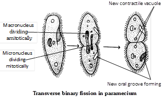
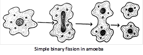 (b) Transverse binary fission : If the plane of cytoplasmic division coincides with the transverse axis of the individual, the fission is termed transverse binary division. Example - Paramecium and Planaria.
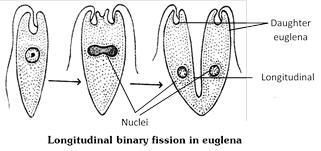
 (c) Longitudinal binary fission : If the plane of cytoplasmic division concides with the longitudinal axis of the individual. This kind of fission is designated as longitudinal binary fission. Example Euglena and vorticella.
 Binary fission involves mitosis only and consequently, the resultant offspring's are genetically identical to the parent and each other.
(ii) Multiple fission : Multiple fission is the division of the parent into many small daughter individuals simultaneously. Examples – Multiple fission occurs in many protozoans such as Plasmodium, more...
Binary fission involves mitosis only and consequently, the resultant offspring's are genetically identical to the parent and each other.
(ii) Multiple fission : Multiple fission is the division of the parent into many small daughter individuals simultaneously. Examples – Multiple fission occurs in many protozoans such as Plasmodium, more...
Definition : Ageing is the slow deterioration in the structure and function of body cells tissues and organs of an animal and starts after the adulthood.
Gerontology : The field of developmental biology that deal with the process and problems of ageing is known as gerontology – (Gr. geron = old man; logos = discourse). The scientists involved in the science of ageing are called gerontologist.
Life cycle and life span : In all metazoan animals, the life cycle includes two developmental period; embryonic period (pre-natal developmental period) which extends from zygote to offspring till hatching or birth, and post embryonic period (post-natal developmental period)- which includes growth, adulthood, reproduction, ageing. Thus, the life cycle comprises five main events : birth, growth, maturity, old age and death, that follow in the sequence named. Maximum life span is the maximum number of years survived by any member of a species, while average life span is the number of years survived by members of a population. Life expectancy is the age at which half the population still survives.
Life span in different organisms
|