
 (5) Vital index : The percentage ratio of natality over mortality is known as vital index i.e. natality / mortality \[\times 100.\] It determines the growth of a population.
(6) Immigration : It is permanent entry of additional person into the existing population of a country or region from out side. Example; Many Nepalese and Chinese come to settle in India.
(7) Emigration : It is the permanent departure of some persons from the existing population of a region to a different state or a foreign country. Example; Many Indians go to Western more...
(5) Vital index : The percentage ratio of natality over mortality is known as vital index i.e. natality / mortality \[\times 100.\] It determines the growth of a population.
(6) Immigration : It is permanent entry of additional person into the existing population of a country or region from out side. Example; Many Nepalese and Chinese come to settle in India.
(7) Emigration : It is the permanent departure of some persons from the existing population of a region to a different state or a foreign country. Example; Many Indians go to Western more... 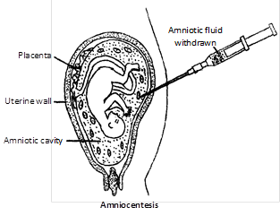 (3) A small amount of amniotic fluid is withdrawn. It contains foetal skin cells and a number of proteins, especially enzymes. The cells can be cultured in vitro for further examination.
Significance
(1) Sex determination : The somatic cells of foetal skin drawn with the amniotic fluid are stained to determine the presence of sex chromatin (barr body). Presence of barr body indicates that the developing foetus in female as female is with 2 X-chromosomes out of which one X-chromosome is active, while other X-chromosome is heterochromatised into a darkly stained barr body.
(2) Congential disease : By Karyotypic studies of somatic cells, abnormalities due to changes in chromosome number like Down's syndrome, Turner's syndrome, Klinefelter's syndrome etc. can be determined.
(3) Metabolic disorder : By the enzyme analysis of amniotic fluid, different types of inborn metabolic disorders like phenylketonuria, alcaptonuria etc. can be detected. These inborn errors are caused by the absence or inactivity of specific enzymes due to gene mutations. So with the help of amniocentesis, if it is confirmed that the child is likely to suffer from some incurable, congenital defect, the mother can go for abortion.
(4) Drawback : However, these days, the amniocentesis is being misused also. Mothers even get their normal foetus aborted if it is a female. This is just equivalent to killing of a normal child. So Govt. of India enforced the Pre-natal Diagnostic Techniques (Regulation and Prevention of Misuse) Act, 1994, since January 1, 1994 under which all genetic counselling centres and laboratories are required to apply for registration. The violation of this Act can bring a fine of Rs. 50,000 and imprisonment for two years. The doctor's registration is also cancelled till the complaint is disposed of.
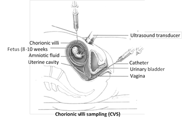
(5) Chorionic Villus Sampling (CVS) : Amniocentesis is possible without a chance of injuring the foetus with the needle only after the sixteenth week of pregnancy. At this time, abortion is not safe. A new technique, named Chorionic Villus Sampling (CVS), can be done during the eighth to tenth week of pregnancy when abortion is safe for the woman. For CVS, cells are sucked into a catheter passed through the cervix. CVS technique provides a mass of rapidly dividing foetal cells, thus facilitating the examination of chromosomal disorders.
(3) A small amount of amniotic fluid is withdrawn. It contains foetal skin cells and a number of proteins, especially enzymes. The cells can be cultured in vitro for further examination.
Significance
(1) Sex determination : The somatic cells of foetal skin drawn with the amniotic fluid are stained to determine the presence of sex chromatin (barr body). Presence of barr body indicates that the developing foetus in female as female is with 2 X-chromosomes out of which one X-chromosome is active, while other X-chromosome is heterochromatised into a darkly stained barr body.
(2) Congential disease : By Karyotypic studies of somatic cells, abnormalities due to changes in chromosome number like Down's syndrome, Turner's syndrome, Klinefelter's syndrome etc. can be determined.
(3) Metabolic disorder : By the enzyme analysis of amniotic fluid, different types of inborn metabolic disorders like phenylketonuria, alcaptonuria etc. can be detected. These inborn errors are caused by the absence or inactivity of specific enzymes due to gene mutations. So with the help of amniocentesis, if it is confirmed that the child is likely to suffer from some incurable, congenital defect, the mother can go for abortion.
(4) Drawback : However, these days, the amniocentesis is being misused also. Mothers even get their normal foetus aborted if it is a female. This is just equivalent to killing of a normal child. So Govt. of India enforced the Pre-natal Diagnostic Techniques (Regulation and Prevention of Misuse) Act, 1994, since January 1, 1994 under which all genetic counselling centres and laboratories are required to apply for registration. The violation of this Act can bring a fine of Rs. 50,000 and imprisonment for two years. The doctor's registration is also cancelled till the complaint is disposed of.
(5) Chorionic Villus Sampling (CVS) : Amniocentesis is possible without a chance of injuring the foetus with the needle only after the sixteenth week of pregnancy. At this time, abortion is not safe. A new technique, named Chorionic Villus Sampling (CVS), can be done during the eighth to tenth week of pregnancy when abortion is safe for the woman. For CVS, cells are sucked into a catheter passed through the cervix. CVS technique provides a mass of rapidly dividing foetal cells, thus facilitating the examination of chromosomal disorders.
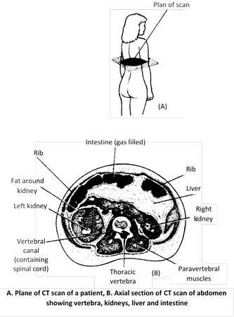 (3) Positron Emission Tomographic Scanning (PET)
(i) PET scanning was developed by Louis Sokoloff of USA in 1985.
(ii) PET is a diagnostic technique based on detection of positrons (positively charged electrons) emitted by radio isotopes such as carbon 11, nitrogen 13 or oxygen 15 generated by the cyclotron.
(iii) These radio isotopes are then incorporated by chemical methods into biological molecules such as glucose, amino acids, carbon dioxide and ammonia.
(iv) These positron emitting compounds are injected into the blood-stream and are taken up in greater concentration by areas of tissues that are more metabolically active.
(v) PET scanning provides three- dimensional images that reflect the metabolic and chemical activity of tissues beings studied.
(vi) PET scanning is particularly valuable for measurement of regional cerebral blood volume, blood flow, metabolic rates of glucose and oxygen in humans.
(vii) PET scanning is used for detecting tumours, for locating the origin of epileptic activity within brain more...
(3) Positron Emission Tomographic Scanning (PET)
(i) PET scanning was developed by Louis Sokoloff of USA in 1985.
(ii) PET is a diagnostic technique based on detection of positrons (positively charged electrons) emitted by radio isotopes such as carbon 11, nitrogen 13 or oxygen 15 generated by the cyclotron.
(iii) These radio isotopes are then incorporated by chemical methods into biological molecules such as glucose, amino acids, carbon dioxide and ammonia.
(iv) These positron emitting compounds are injected into the blood-stream and are taken up in greater concentration by areas of tissues that are more metabolically active.
(v) PET scanning provides three- dimensional images that reflect the metabolic and chemical activity of tissues beings studied.
(vi) PET scanning is particularly valuable for measurement of regional cerebral blood volume, blood flow, metabolic rates of glucose and oxygen in humans.
(vii) PET scanning is used for detecting tumours, for locating the origin of epileptic activity within brain more... 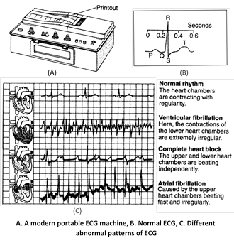 (3) Electroencephalograph
(i) The electrical activity of the exposed animal brain was discovered by Satton in 1875.
(ii) Hans Berger (1929) was the first to record Electro-Encephalo-Gram (EEG).
(iii) Electroencephalography is done by attaching a number of small electrodes to the scalp.
(iv) The electrodes are connected to an instrument that measures the brain's impulses in microvolts and amplifies them for recording purposes.
(v) Electroencephalography is painless, produces no side-effects and to record it takes about 45 minutes.
(vi) An EEG records the minute electrical impulses produced by the activity of brain.
(vii) EEG is useful to find out whether the person is alert, awake or asleep.
(viii) EEG can help in diagnosing certain conditions such as epilepsy, encephalitis, dementia and brain tumour.
(ix) Electroencephalography can also be used to monitor the condition of patients during surgery and to assess the depth of anaesthesia.
(x) EEG is also used as a test for brain death.
(xi) The weaker magnetic fields from the brain can be studied with the help of SQUID (Super conducting Quantum Interference Device).
(xii) Magnetoencephalography (MET) is useful for studying the disease associated with the brain and spinal cord.
(3) Electroencephalograph
(i) The electrical activity of the exposed animal brain was discovered by Satton in 1875.
(ii) Hans Berger (1929) was the first to record Electro-Encephalo-Gram (EEG).
(iii) Electroencephalography is done by attaching a number of small electrodes to the scalp.
(iv) The electrodes are connected to an instrument that measures the brain's impulses in microvolts and amplifies them for recording purposes.
(v) Electroencephalography is painless, produces no side-effects and to record it takes about 45 minutes.
(vi) An EEG records the minute electrical impulses produced by the activity of brain.
(vii) EEG is useful to find out whether the person is alert, awake or asleep.
(viii) EEG can help in diagnosing certain conditions such as epilepsy, encephalitis, dementia and brain tumour.
(ix) Electroencephalography can also be used to monitor the condition of patients during surgery and to assess the depth of anaesthesia.
(x) EEG is also used as a test for brain death.
(xi) The weaker magnetic fields from the brain can be studied with the help of SQUID (Super conducting Quantum Interference Device).
(xii) Magnetoencephalography (MET) is useful for studying the disease associated with the brain and spinal cord. 





You need to login to perform this action.
You will be redirected in
3 sec